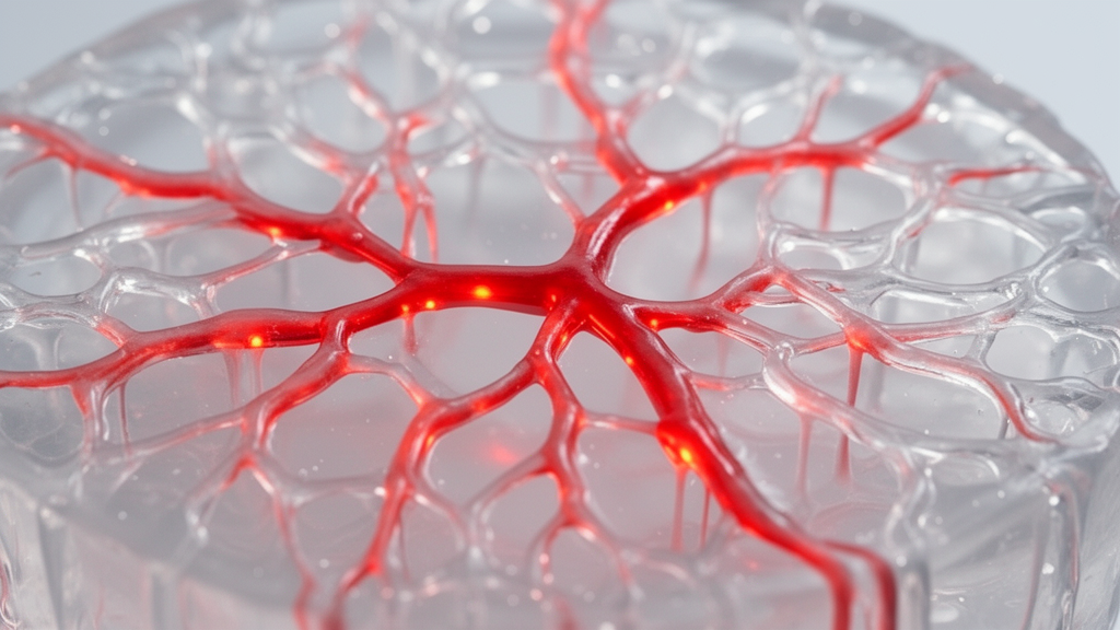
Bioengineers have published an end-to-end laboratory protocol detailing a diffusion-driven three-dimensional bioprinting methodology, termed Gelation of Uniform Interfacial Diffusant in Embedded 3D Printing, to fabricate self-supporting, perfusable vascular networks with tunable dimensions. Published in the journal BMC Methods, the protocol resolves a persistent challenge in sacrificial ink extrusion, where constructing hollow tubular structures with distinct, self-supporting walls has historically proven difficult. In tissue engineering, functional vasculature is vital for sustaining cell and tissue viability by enabling transport of oxygen, signaling molecules, nutrients, and waste. The GUIDE-3DP technique utilizes the interfacial transport of chemical crosslink initiators from an extruded sacrificial ink into a surrounding hydrogel precursor bath. When diffusing initiators reach a critical concentration threshold within the surrounding matrix, localized gelation is triggered around the strand, generating hollow, interconnected architectures that replicate complex physiological geometries.
The bioprinting technique relies on Fickian diffusion to control crosslinking kinetics and spatial boundaries at the bioink interface. Embedded printing methods extrude bioinks into support baths to sustain soft materials and non-planar geometries, while sacrificial ink approaches deposit removable filaments into bulk matrices that are cleared to leave open channels. GUIDE-3DP combines these approaches by transforming the sacrificial filament into a localized initiator delivery vehicle rather than a temporary template. Prior to printing, finite element modeling can simulate concentration gradients and crosslinking dynamics across complex branched geometries, while empirical diffusion parameters can be evaluated through fluorescence imaging and fluorescence recovery after photobleaching. By establishing a predictable time-dependent diffusion gradient outward from the extruded ink into the bulk precursor, researchers can program the crosslinking of the vessel wall shell, providing a reliable workflow for producing branching networks.
The initial phase of the protocol involves designing freeform vascular geometries within computer-aided modeling software, specifically Rhinoceros 3D, and translating those paths into G-code instructions. Print designs can be derived from anatomical reference data in repositories like NIH3D, the Vascular Model Repository, or BioRender diagrams. Within the modeling environment, researchers trace network paths using control point curve tools, deliberately introducing slight geometric overlaps between intersecting segments to prevent discontinuous inner lumens during extrusion. The continuous paths are subsequently segmented using the divide function to yield coordinate points spaced at uniform intervals, such as 0.5 millimeters, and exported into text files. Because filament diameter is governed by point spacing intervals and extrusion values, operators select extrusion values typically between 0.005 and 0.02 millimeters of extrusion movement per point to modulate ink deposition volume.
Following coordinate extraction, the numerical data are imported into spreadsheet software such as Excel to generate G-code lines following standard linear motion command syntax. The toolpath trajectories are simulated in G-code editing environments like CAMotics to verify travel directions and geometric fidelity. Crucial machine preparation codes are appended before toolpaths begin, including relative extrusion mode M83, baseline coordinate setting G92, and print speed defined in millimeters per minute. To prevent inadvertent material leakage and trailing artifacts between non-contiguous vascular branches, the authors specify that the printhead re-trace existing paths in reverse with extrusion set to zero, rather than traversing open bath volume where dragging sacrificial ink would disrupt the geometry. Finally, vertical nozzle retract movements are appended to lift the nozzle above the support bath, preserving the fragile, uncrosslinked structure.
To establish a cell-compatible support bath precursor, the protocol outlines a large-scale synthesis method for gelatin methacryloyl derived from cold water fish skin gelatin. Unlike porcine gelatin, cold water fish gelatin remains liquid at room temperature, facilitating smooth translation of the print nozzle through the bath. Synthesis begins by dissolving cold water fish gelatin at 20 percent weight per volume in 1 molar carbonate-bicarbonate buffer, adjusting to pH 10 using 5 molar sodium hydroxide, and heating to 70 degrees Celsius. Methacrylic anhydride is added at 0.0833 milliliters per gram of gelatin under continuous stirring for two hours. The functionalized GelMA is purified through either five days of dialysis using a 10 kilodalton molecular weight cutoff membrane or ethanol precipitation. Proton nuclear magnetic resonance spectroscopy confirmed a degree of methacrylation of 70.7 plus or minus 5.2 percent across four synthesis batches, calculated from normalized lysine integral signals relative to aromatic amino acid signals.
Preparing the embedded matrix involves combining the gel precursor with a rheological modifier to yield a stable, shear-thinning, and self-healing support bath. The authors describe formulations using either GelMA at 10 to 20 weight percent or alginate at 2 weight percent, blended with Aristoflex AVC as a viscosity modifier at 2 weight percent. For a standard GelMA formulation, 40 weight percent GelMA stock is mixed one-to-one with 4 weight percent Aristoflex AVC stock, optionally supplemented with 0.5 to 1 weight percent fibrinogen. To eliminate air bubbles, dishes containing 8 grams of support material per well in six-well plates are centrifuged at 4,000 revolutions per minute for five minutes. Concurrently, sacrificial inks are formulated using gelatin microparticle slurries at 100 milligrams per milliliter or Pluronic F-127 at 26 weight percent, loaded with crosslink initiators including LAP at 1 to 5 millimolar, Eosin Y disodium salt at 0.025 to 0.1 millimolar, or calcium chloride at 50 to 100 millimolar.
To enable multi-material printing, the protocol provides an optional calibration routine for dual syringe pump extruders mounted on an open-source bioprinter. Spatial alignment across X, Y, and Z axes is achieved through a bed-mounted calibration camera or a precision-machined reference block. Using the camera method, primary nozzle Tool 0 is focused and registered as the origin using G92 and G10 commands, after which secondary nozzle Tool 1 is centered and its relative displacement is determined using coordinate queries. In the block method, the operator aligns each nozzle sequentially against the orthogonal X, Y, and Z faces of a reference cube, verifying contact by tapping the needle with forceps. Relative tool offset parameters are stored in firmware via G10 commands, and custom tool-change routines incorporate automated vertical lift distances of 10 millimeters to clear support bath boundaries and eliminate collision risks during extruder transitions.
During physical fabrication, the loaded print syringe fitted with a 27-gauge blunt needle is positioned within the support bath, executing the programmed toolpath at designated extrusion velocities. Following extrusion, constructs undergo incubation at room temperature for 5 to 15 minutes, allowing crosslink initiators to diffuse outward into the adjacent GelMA or alginate matrix. Light-activated crosslinking is then initiated, utilizing ultraviolet exposure for 5 minutes when LAP is the initiator, or white-light illumination for 10 minutes when Eosin Y is employed alongside triethanolamine and 1-vinyl-2-pyrrolidinone co-initiators. Following crosslinking, the hydrogel construct is extracted from the uncrosslinked support bath, washed in saline buffer, and transferred into cell culture medium at 37 degrees Celsius. At this temperature, the sacrificial gelatin microparticle core melts, closed channel ends are excised with surgical tools, and warm medium irrigation clears the hollow vascular lumen.
The protocol details two distinct methodologies for endothelializing hollow lumens, utilizing either post-fabrication fluid perfusion or direct cell-laden sacrificial ink deposition. In the direct printing approach, endothelial cells are suspended within the gelatin microparticle ink at a density of ten million cells per milliliter adjusted to neutral pH. Following crosslinking and bath removal, the construct is incubated at 37 degrees Celsius for 10 minutes, allowing the sacrificial matrix to liquefy while suspended endothelial cells sediment onto the lower luminal wall. The construct is inverted and incubated for another 10 minutes, facilitating uniform cellular attachment across the opposing luminal surface before residual liquid gelatin is gently flushed out. Quantitative cell counter assessments indicated high retention rates between 70 and 90 percent of initial cell populations, while immunofluorescence imaging confirmed confluent endothelial monolayers exhibiting localized VE-cadherin expression at cell-cell junctions after five days of culture.
Experimental evaluations demonstrated that the GUIDE-3DP protocol confers independent control over internal lumen diameters and external channel wall thicknesses by adjusting specific operational parameters. Increasing sacrificial ink extrusion rates from 0.6 to 2 microliters per second, or shifting from a 32-gauge to a 27-gauge needle at a fixed print speed of 180 millimeters per minute, systematically produced larger internal lumen diameters. Printing across upward, downward, and horizontal trajectories yielded nearly identical internal dimensions, although horizontal paths exhibited slightly higher variance due to minor lateral filament spreading within the support bath. Channel wall thickness was directly modulated by varying crosslink initiator concentration and interfacial diffusion duration, showing that longer diffusion periods and higher initiator concentrations expanded outer wall dimensions. Demonstrating versatility across retinal vasculature models, hierarchical branching networks, and dual-material capillary constructs, the authors note that the method is adaptable to engineer other hollow tissue analogs, including intestinal epithelia, fallopian tubes, and airway channels.
Subject of Research: Diffusion-based embedded 3D bioprinting of perfusable vascular networks
Article Title: A diffusion-based 3D printing strategy to fabricate self-supporting, perfusable networks
Article References: Ramos Mejia, D., Cai, B., Iranzo, S. C., Perez, A., Tan, Y. L., Lee, S., & Heilshorn, S. C. (2026). A diffusion-based 3D printing strategy to fabricate self-supporting, perfusable networks. BMC Methods, 3(1), Article 8. https://doi.org/10.1186/s44330-026-00059-6
Image Credits: AI Generated
DOI: 10.1186/s44330-026-00059-6
Keywords: 3D bioprinting, tissue engineering, vascular networks, gelatin methacryloyl, sacrificial ink, GUIDE-3DP, biofabrication, endothelialization, diffusion-based, printing, strategy, fabricate
News Source: Gregory Coleman. (November 12, 2026). Diffusion-Based 3D Bioprinting Strategy Yields Self-Supporting Perfusable Vascular Networks. Scienmag.