Information technology can make life easier in many areas – including research. In medicine, for example, it is still standard practice to evaluate microscopy images of tissue sections by hand. This is used, for example, to assess how many cancer cells are in a lymph node.
“You often sit in a dark room for hours counting the cells on an image captured with a fluorescence microscope. That costs an incredible amount of valuable time,” says Philipp Sodmann, who works in cardiac research at the University Hospital of Würzburg in Bavaria, Germany.
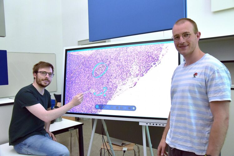
But now a new horizon is opening up for the life sciences: The new digital tool deepflash2 makes the analysis of microscopy images much easier. deepflash2 is freely available and based on machine learning methods.
Jury emphasizes on quality aspect
Matthias Griebel from the Chair of Information Systems and Business Analytics at the University of Würzburg developed the tool as part of his doctorate. The tool formed the foundation of the solution he developed together with medical scientist Philipp Sodmann for an international data science competition. In this competition, the team of the two from Würzburg was successful: in May 2021, it received the innovation award endowed with 10,000 US dollars and a gold medal from the online platform Kaggle.
The top-class jury with experts from medicine, biology, and artificial intelligence (AI) attested deepflash2 another quality: the program also recognizes ambiguities.
“In biology, not everything is black or white,” explains Matthias Griebel. It is not uncommon for researchers to doubt whether cells they see in a tissue section are still functional. In such cases, deepflash2 points this out: People have to look at this again! This is what makes the tool particularly innovative, according to the jury members.
Freely available for researchers
deepflash2 is still an insider’s tip for researchers involved in bio-image analysis. However, Matthias Griebel wants to use the excellent results in the data science competition as an opportunity to promote his tool.
Since it is an open-source tool, other researchers can use it free of charge in their browser or install it on their computer. In the meantime, Griebel is already working on further improving deepflash2 using the findings from the competition.
deepflash2 on Github: https:/
Applicable even without AI knowledge
Griebel, who studied business information systems, is doing his doctorate under Professor Christoph Flath. During the development of deepflash2, he attached great importance to the fact that researchers without AI expertise can also use the tool without any problems.
Users from medicine and life sciences must not understand the complicated processes behind the scenes. For them, according to Griebel, the most important thing is to make bio-image analysis faster and at the same time more reliable. To achieve this, an artificial neural network must be intensively trained using extensive data sets, says the Würzburg scientist.
Decisions are made by humans
In the end, however, it is humans who draw a conclusion from the images. This should reassure all those who fear that artificial intelligence will decide the weal and woe of medicine in the future. Philipp Sodmann emphasizes that this is not the case and will certainly never become the case soon.
Sodmann appeals to recognize the manifold possibilities of AI. The data science competition, for example, took place against the backdrop of the “Human BioMolecular Atlas Program” project launched in 2016. Its goal is to map and characterize every single one of the approximately 37 trillion human cells. This would be impossible without AI.
Prize for the best presentation
A total of around 1,200 teams from more than 50 countries submitted solutions for the Kaggle data science competition. Matthias Griebel and Philipp Sodmann landed in 10th place.
“Whereby the first places were decided in a neck-and-neck race,” says Griebel. The presentation of the project in front of an international audience was also exciting for him and his colleague. The two Würzburgers came out on top again: they also won the prize for the best presentation, in addition to the gold medal and the innovation prize.
Suitable for different clinical pictures
Matthias Griebel does not want to research in an ivory tower. It is important to him to develop tools that will ultimately help people. And perhaps even save lives.
If microscopy images can be evaluated faster and more reliably, a diagnosis can also be made more quickly. And this is true for very different diseases. Because the deepflash2 program is trainable, it can learn, for example, to recognize different functional tissue units. Thus, with the help of machine learning, the algorithm can be taught to identify the insulin-producing cells of the pancreas on an image.
###
Media Contact
Matthias Griebel
[email protected]
Original Source
https:/


