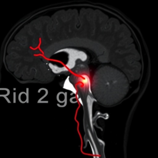
In a groundbreaking advancement bridging forensic pathology and cutting-edge imaging technology, a new study unveils a meticulously detailed method for identifying vertebral artery injuries resulting from stab wounds. This innovative approach harnesses the power of contrast-enhanced fluoroscopy coupled with micro-computed tomography (micro-CT), allowing for unprecedented ex-situ visualization and analysis of vascular trauma that was previously difficult to thoroughly assess. The implications of this technique extend far beyond academic curiosity, offering profound potential for forensic investigations, clinical diagnostics, and even surgical planning.
Vertebral artery injuries, especially those inflicted by penetrating trauma such as stab wounds, present distinct challenges in forensic examinations due to the artery’s deep anatomical location and its complex relationship with surrounding osseous and soft tissues. Traditional methods of injury detection often rely upon gross dissection and standard radiological techniques that may fail to provide the resolution necessary to distinguish intricate injury patterns or to map the extent of arterial compromise fully. This novel approach fills a critical gap by exploiting the unique strengths of both contrast-enhanced fluoroscopy and the ultra-high resolution capabilities of micro-CT.
At the core of this methodology lies the principle of ex-situ examination, wherein the vertebral artery, extracted intact or closely associated with surrounding bony structures, is subjected to advanced imaging outside the body. This process eliminates motion artifacts and physiological interferences, ensuring that the resulting images are stable and highly detailed. Contrast agents are introduced into the arterial lumen, significantly enhancing the visibility of the vascular pathways and potential sites of injury. Such enhancement is vital for discerning subtle disruptions in vessel integrity that could be missed during in situ imaging scenarios.
Fluoroscopy, renowned for its real-time imaging capabilities, serves as the initial screening tool in this workflow. The live imaging allows researchers and forensic experts to observe the flow of contrast medium through the vertebral artery, promptly identifying occlusions, leaks, or stenotic segments indicative of trauma. However, fluoroscopy’s relatively modest spatial resolution demands a supplementary technique for more granular scrutiny. This is where micro-CT steps in, delivering microscopic-level cross-sectional images that reveal the three-dimensional architecture of the vascular injury with extraordinary clarity.
Utilizing micro-CT involves rotating the specimen while capturing thousands of X-ray images, which are computationally reconstructed into a detailed volumetric dataset. In this way, microscopic features such as intimal tears, intraluminal thrombi, or dissections of the vessel wall become vividly visible. The digital models generated also permit virtual dissections and explorations from multiple angles, providing forensic experts with comprehensive assessments often difficult to achieve through physical examination alone. This digital preservation also supports prompt reanalysis and sharing across diverse investigatory teams.
The authors of the study emphasize the distinct advantage this combination of techniques holds in forensic casework. Where autopsy findings can be confounded by decomposition, surgical intervention, or complex trauma patterns, an ex-situ imaging protocol offers a consistent framework for standardizing injury assessment. This reproducibility is crucial for judicial contexts, where detailed, incontrovertible documentation of traumatic vascular injuries can influence legal determinations and expert testimonies.
Beyond forensic applications, the implications for clinical practice are significant. Accurately mapping vertebral artery injuries is critically important for neurosurgeons and trauma physicians managing patients with cervical penetrating injuries. The novel imaging approach, though currently primarily deployed ex situ, paves the way for less invasive, high-resolution diagnostic tools that could in the future guide in vivo interventions and preoperative planning, potentially improving patient outcomes.
The detailed images obtained are more than mere clinical aids—they reveal the biomechanical interplay between penetrating weapons and the delicate vascular structures nestled within the cervical spine. Understanding these injury mechanisms at a microscopic scale enlightens researchers about the forces required to disrupt vertebral arteries, informs the design of protective gear, and guides the development of more sophisticated forensic reconstructions of violent events.
Critically, the study leveraged state-of-the-art contrast agents optimized for high radiodensity and vascular compatibility, ensuring that the enhancement was both sharp and artifact-free. This careful chemical design was essential for achieving the clarity necessary to differentiate between true vascular injury and postmortem artifacts, a perennial challenge in forensic imaging. Moreover, by preserving tissue integrity during imaging, the specimens remain amenable to subsequent histological or molecular analyses if needed.
In addition to technical innovation, the research highlights a broader methodological paradigm shift in forensic science—one that embraces multidisciplinary collaboration invested in technological synergy. Radiologists, pathologists, chemists, and imaging specialists came together to refine protocols, calibrate imaging parameters, and validate findings against traditional autopsy results. This convergence underscores a holistic approach to solving complex forensic puzzles that transcends singular disciplinary perspectives.
Furthermore, the accessibility of micro-CT technology is rapidly increasing, with scanners becoming more affordable and user-friendly for institutions beyond specialized research facilities. This democratization means that detailed vascular injury assessments could become a routine part of forensic investigations globally, heralding higher standards in trauma documentation and judicial processes.
The visual documentation generated has also garnered significant attention for its educational value. Dynamic fluoroscopy videos and three-dimensional reconstructions provide compelling teaching tools for medical students, forensic specialists, and even law enforcement professionals, allowing them to visualize unseen pathological processes with remarkable clarity. These visual aids demystify complex injury patterns and enhance understanding of trauma biomechanics in ways textbooks alone cannot convey.
This novel methodological framework additionally establishes a robust foundation for future studies exploring other vascular territories involved in penetrating trauma. Arteries such as the carotid, subclavian, or even intracranial vessels could be similarly examined utilizing tailored ex-situ imaging protocols, enhancing our collective knowledge of traumatic vascular pathology and expanding forensic diagnostic capabilities.
The interdisciplinary nature and innovative imaging fusion presented in this research illustrate how technological advances continue to push beyond the boundaries of classical forensic medicine. As imaging tools evolve toward greater resolution, faster acquisition, and multi-contrast capabilities, the feasibility of fully non-invasive or minimally invasive postmortem vascular examinations appears increasingly attainable.
In conclusion, this novel imaging protocol combining contrast-enhanced fluoroscopy with micro-CT ex situ represents a paradigm shift in forensic trauma evaluation—delivering unprecedented detail, accuracy, and reproducibility in the assessment of vertebral artery injuries caused by stab wounds. It paves the way for enhanced forensic reconstructions, improved clinical diagnostics, and a deeper biomechanical understanding of vascular trauma, signifying a remarkable step forward at the intersection of technology and forensic science.
Subject of Research: Identification of vertebral artery injuries caused by stab wounds using advanced imaging techniques.
Article Title: Ex-situ identification of vertebral artery injuries from stab wounds through contrast-enhanced fluoroscopy and micro-CT.
Article References:
Secco, L., Franchetti, G., Viel, G. et al. Ex-situ identification of vertebral artery injuries from stab wounds through contrast-enhanced fluoroscopy and micro-CT. Int J Legal Med (2025). https://doi.org/10.1007/s00414-025-03608-w
Tags: clinical diagnostics for vascular injuriescontrast-enhanced fluoroscopy applicationsex-situ examination in pathologyforensic investigation techniquesforensic pathology advancementsintricate injury pattern analysismicro-computed tomography in forensicspenetrating trauma assessmentstab wound detection techniquessurgical planning with advanced imagingvascular trauma imaging methodsvertebral artery injuries


