In the war against cancer, the first step towards treating them is an accurate diagnosis of the problem. A technique that helps plan a better course of treatment is a biopsy. In recent years, liquid biopsies, which enable the detection of diseases via blood or other bodily fluids instead of solid tissues, have gained popularity over surgical biopsies owing to their non-invasive and straightforward nature.

Credit: Nobuyuki Futai from SIT, Japan
In the war against cancer, the first step towards treating them is an accurate diagnosis of the problem. A technique that helps plan a better course of treatment is a biopsy. In recent years, liquid biopsies, which enable the detection of diseases via blood or other bodily fluids instead of solid tissues, have gained popularity over surgical biopsies owing to their non-invasive and straightforward nature.
Liquid biopsies primarily target a molecular marker called “cell-free DNA” (cfDNA), which provides information about the presence of pathogenic DNAs in a sample. To carry out any form of analysis, cfDNA has to be extracted and purified, a challenging task owing to their low abundance. A gold-standard cfDNA purification method called “solid-phase extraction” relies on the DNA’s affinity towards a solid phase. However, it fails to yield DNA fragments with less than 200 base pairs (bp)—fundamental units of DNA. But, why is sensitivity towards smaller fragments a necessity? It is said that circulating tumor DNA (ctDNA) or pathogenic DNAs are typically smaller than cfDNA. Therefore, sensitivity towards DNA fragments smaller than 200 bp allows for better detection of diseases.
Newer techniques such as liquid phase extraction (LPE), isotachophoresis (ITP), and electrokinetic trapping of DNA can facilitate such size-independent extractions. However, LPE is very labor-intensive and time-consuming. ITP and electrokinetic trapping, despite their excellent abilities in providing quick and automated extraction and detection of pathogenic DNA e.g., M. tuberculosis (MTB), have not been explored for the selective purification of short cfDNA fragments.
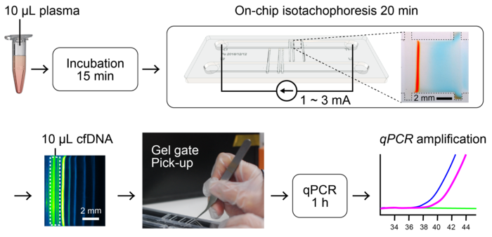
Now, in a recent study published in Analytica Chimica Acta, researchers from Japan and USA have demonstrated a novel extraction system that combines the powers of both ITP and electrokinetic trapping. Led by Professor Nobuyuki Futai from Shibaura Institute of Technology (SIT), Japan, the team designed an open microfluidic system that uses transient ITP to detect MTB from human plasma samples. Prof. Futai explains, “The millimeter-scale fluidic device we developed consists of movable gel gates that allow precise extraction of separate species. It uses ITP for the purification of DNA, and the purified DNA can be easily extracted as a PCR-ready gel strip.”
To test the efficacy of the design, the team used the device to purify and enrich MTB-genomic DNA fragments from spiked human plasma. The fluidic system showed a high recovery rate, precise separation, and sensitivity towards short cfDNA fragments of 100-200 bp. It was also able to purify MTB DNA for further qPCR analysis. Further investigation into its separation abilities revealed that treatment of the plasma with the enzyme proteinase K generated plasma peptides. These peptides acted as endogenous spacer molecules and improved the resolution of the extraction method.
The designed reconfigurable open microchannel device creates a versatile sample preparation platform for analysis techniques such as PCR and deep sequencing. The team believes that their findings could be used to develop advanced purification systems for recovering nucleic acids from plasma or serum.
“Most biopsy sample preparation techniques use marker dyes during DNA purification, which often leads to contamination and decrease in the qPCR signal level. The specific chemistries and sieving effects created by our movable gate design not only ensures the excellent recovery of purified DNA but also eliminates the need for marker dyes,” comments Prof. Futai. “This would, for instance, enable accurate diagnosis of diseases and infections from a small amount of blood sample.”
Those, certainly, are consequences to look forward to!
***
Reference
DOI: https://doi.org/10.1016/j.aca.2022.339435
About Shibaura Institute of Technology (SIT), Japan
Shibaura Institute of Technology (SIT) is a private university with campuses in Tokyo and Saitama. Since the establishment of its predecessor, Tokyo Higher School of Industry and Commerce, in 1927, it has maintained “learning through practice” as its philosophy in the education of engineers. SIT was the only private science and engineering university selected for the Top Global University Project sponsored by the Ministry of Education, Culture, Sports, Science and Technology and will receive support from the ministry for 10 years starting from the 2014 academic year. Its motto, “Nurturing engineers who learn from society and contribute to society,” reflects its mission of fostering scientists and engineers who can contribute to the sustainable growth of the world by exposing their over 8,000 students to culturally diverse environments, where they learn to cope, collaborate, and relate with fellow students from around the world.
Website: https://www.shibaura-it.ac.jp/en/
About Professor Nobuyuki Futai from SIT, Japan
Dr. Nobuyuki Futai is currently a professor at the Department of Mechanical Engineering, Shibaura Institute of Technology. Dr. Futai’s expertise lies in the field of Bioengineering and Mechanical Engineering. His current research endeavor includes reconfigurable microfluidic devices useful in cell culture and biology. Along with his team, Dr. Futai also researches mobile viral culture devices and systems to collect and analyze marine microplastics.
Funding Information
This study was supported by JSPS KAKENHI Grant Number 16K01374.
Journal
Analytica Chimica Acta
DOI
10.1016/j.aca.2022.339435
Method of Research
Experimental study
Subject of Research
Human tissue samples
Article Title
A modular and reconfigurable open-channel gated device for the electrokinetic extraction of cell-free DNA assays
Article Publication Date
8-Jan-2022
COI Statement
The authors declare that they have no known competing financial interests or personal relationships that could have appeared to influence the work reported in this paper


