The new technology has been developed jointly by teams headed by Prof Dr Georg Schmitz at the Chair for Medical Engineering at Ruhr-Universität Bochum and by Prof Dr Fabian Kiessling at the Institute for Experimental Molecular Imaging at the University Hospital Aachen. They published their report in the journal Nature Communications from April 18, 2018.
Monitoring microbubbles on their path through the body
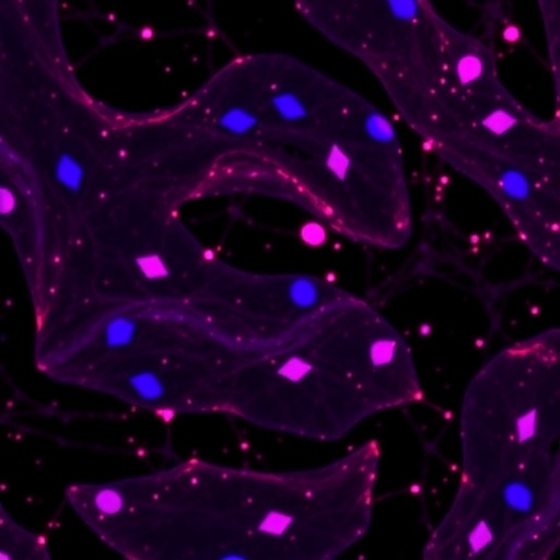
The new technology called "Motion Model Ultrasound Localization Microscopy" is based on contrast medium-enhanced ultrasound images. Microbubbles are administered to patients as contrast agents: gas bubbles no larger than one micrometre that travel through the body in the bloodstream. In ultrasound images, they appear as shapeless white blobs. "Once the centre of each of these blobs has been identified, it's possible to determine the location of individual bubbles," explains Georg Schmitz.
Each bubble is given a name
Using algorithms originally developed for radar technology, the research team successfully monitored the motion of individual microbubbles. "We are currently attempting to teach the computer something that our eyes are able to do: namely read movement in a sequence of images in which a dot appears in different locations," says Schmitz. To this end, the researchers gave each bubble a name. Thus, they were able to track their paths through the vascular system and count them in the process.
Resolution much higher than mere image resolution
Subsequently, fine vascular networks can be reconstructed based on the motion of the bubbles. The direction and speed of the blood flow can likewise be recorded. The resolution of the images is greatly enhanced: experts refer to the technique as super-resolution imaging.
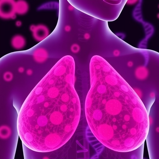
"In the publication, we demonstrated that the synthesis of morphological and functional parameters considerably facilitates the differentiation between tumour types," explains Fabian Kiessling. In the course of their project, they tested the technique in three model cases, including in human subjects. In collaboration with Prof Dr Elmar Stickeler from the Clinic for Gynaecology and Obstetrics at the University Hospital Aachen, the researchers successfully identified how tumour vessels responded to chemotherapy in breast cancer patients.
Monitoring therapy effects
"One reason why this is important is because new therapy approaches aim at manipulating the vascular system of tumours, in order to enhance the therapeutic effect by increasing the concentration of drugs in the tumours," says Fabian Kiessling. One of these approaches is so-called sonoporation. Here, tumours are treated with ultrasound in order to render the vascular walls more permeable to active substances.
"The advantage of our approach is that it can be performed with conventional ultrasound scanners, which have a low frame frequency, with sometimes as few as 15 images per second," points out Georg Schmitz. The research teams have already filed an application for a follow-up project, in the course of which they intend to test the method in large-scale clinical studies.
###
Funding
The project was funded by the German Research Foundation (SCHM1171/4-1, KI1072/11-1)
Original publication
Tatjana Opacic, Stefanie Dencks, Benjamin Theek, Marion Piepenbrock, Dimitri Ackermann, Anne Rix, Twan Lammers, Elmar Stickeler, Stefan Delorme, Georg Schmitz, Fabian Kiessling: Motion model ultrasound localization microscopy for preclinical and clinical multiparametric tumor characterization, in: Nature Communications, 2018, DOI: 10.1038/s41467-018-03973-8, https://www.nature.com/articles/s41467-018-03973-8
Press contact
Prof Dr Georg Schmitz
Chair for Medical Engineering
Faculty of Electrical Engineering and Information Technology
Ruhr-Universität Bochum
Germany
Phone: +49 234 32 27573
Email: [email protected]
Prof Dr Fabian Kiessling
Institute for Experimental Molecular Imaging
University Hospital Aachen
Germany
Phone: +49 241 80 80117
Email: [email protected]
Media Contact
Georg Schmitz
[email protected]
49-234-322-7573
@ruhrunibochum
http://www.ruhr-uni-bochum.de
http://news.rub.de/english/press-releases/2018-04-18-medical-engineering-detailed-images-tumour-vasculature
Related Journal Article
http://dx.doi.org/10.1038/s41467-018-03973-8