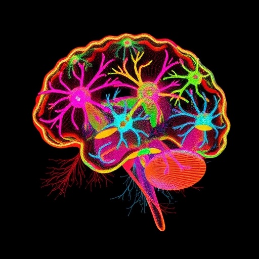
In the intricate landscape of the brain, dendritic arbors form the very architecture through which neurons communicate, compute, and integrate signals. These sprawling, tree-like extensions of neurons serve as the fundamental units for receiving synaptic inputs and orchestrating complex neural computations vital for behavior and cognition. Despite their undeniable importance, comprehensive studies that map the morphology of single neurons across large brain regions have remained an elusive frontier, primarily due to technical limitations. However, a groundbreaking new study by Park, Yan, Zhu, and colleagues promises to transform our understanding of neuronal morphology in the striatum, a crucial brain region implicated in motor control and reward processing. Their innovative approach, aptly termed “dendritome mapping,” meticulously profiles the dendritic structures of genetically defined single neurons in the mouse striatum, revealing spatial patterns and age-related as well as disease-related changes with unprecedented resolution.
The striatum, a subcortical brain structure, is predominantly composed of two types of medium spiny neurons (MSNs), distinguished by their dopamine receptor expression: D1-type and D2-type MSNs. These neurons differ not only in their genetic identity but also in their connectivity and functional roles within basal ganglia circuits. Understanding the distinct morphological features of these neuronal subtypes has profound implications for deciphering how the striatum processes complex information, and how alterations in this processing contribute to neurodegenerative and psychiatric disorders. This ambitious investigation reconstructed 3,762 three-dimensional morphologies of individual D1 and D2 MSNs, which were then accurately mapped to standardized brain reference atlases, enabling high-resolution spatial analysis across the striatum.
One of the major challenges in neuronal morphology studies is capturing the subtle but functionally significant variations in dendritic architectures that are often blurred when relying solely on classical anatomical landmarks. To overcome this, the researchers devised an elegant computational method that divides the striatum into latticed cubic boxes, a spatial grid framework that surpasses the resolution limits of conventional anatomical segmentation. By quantifying a comprehensive set of morphometric parameters within each cubic volume, the team generated compact, high-dimensional morphological signatures—referred to as “eigen-morphs”— encapsulating the unique dendritic features of MSNs within that spatial unit.
The subsequent clustering of these eigen-morphs unveiled six distinct morphological modules, each representing a discrete spatial territory within the striatum. Remarkably, these modules formed contiguous domains, suggesting that the dendritic architecture of MSNs is not a random assortment but rather organized into spatially coherent microcircuits. Each identified module exhibited characteristic dendritic patterns, including variations in branch complexity, length, and orientation, which likely underpin specialized computational functions within the striatal network. Additionally, these morphologically defined territories were shown to receive distinct patterns of corticostriatal innervation, hinting at a direct link between dendritic structure and input specificity that shapes striatal processing.
Beyond merely mapping healthy neuronal morphology, the research team extended their analysis to explore how MSN dendritic architecture evolves with aging and in the context of Huntington’s disease, a devastating neurodegenerative disorder that profoundly affects striatal circuits. Intriguingly, their data revealed a generalized atrophy of dendritic arbors with age in both D1 and D2 MSNs, manifested as reductions in dendritic length and branching complexity. This finding aligns with observed age-associated declines in motor and cognitive functions and suggests that dendritic degradation may be an underlying cellular substrate.
In contrast, the dendritic alterations observed in Huntington’s disease mouse models were far more nuanced and MSN-type specific. The study demonstrated that disease progression induces selective morphological defects, with D2 MSNs showing more pronounced dendritic degeneration in particular striatal subregions. These spatially and cell-type restricted changes provide important clues to the pathogenic mechanisms driving symptom emergence in Huntington’s disease and open new avenues for targeted therapeutic interventions aimed at preserving neuronal structure.
The strength of dendritome mapping lies not only in its technical innovation but also in its integrative systems biology framework, which bridges molecular genetics, high-resolution morphology, and circuit-level analysis. By leveraging genetically encoded neuron-type markers and combining them with high-throughput morphological reconstruction and spatial atlas registration, the researchers established a versatile platform capable of scaling up to other brain regions and neuronal classes. This capability could revolutionize how neuroscientists study cellular diversity and its functional implications in both health and disease.
Technically, the methodology employed involved state-of-the-art imaging techniques, including confocal and two-photon microscopy, followed by meticulous semi-automated 3D reconstruction of neuronal dendrites. Advanced image processing pipelines ensured fidelity and reproducibility across thousands of neurons. The subsequent morphometric quantification entailed calculating an array of structural features—such as dendritic length, branching angles, fractal dimension, and spine density proxies—that together define the morphological phenotype of each neuron. Coupling this with precise stereotactic mapping to a reference atlas enabled the spatial contextualization critical for discovering coherent dendritic modules.
Furthermore, the computational strategies for morphometric data reduction and clustering used dimensionality reduction techniques akin to principal component analysis, hence the term “eigen-morph,” which captures the dominant patterns of variance across the dendritic features. Clustering algorithms then grouped adjacent cubic boxes sharing similar eigen-morph profiles, effectively segmenting the striatum into functionally relevant morphological zones. This scalable computational framework sets a new standard for morphometric neuroanatomy, offering an objective and reproducible means to parse neuronal diversity.
The implications of this research transcend the striatum alone. Since dendritic morphology directly influences neuronal input integration, plasticity, and ultimately circuit dynamics, the dendritome mapping approach provides a quantitative foundation for linking microstructural variability with functional heterogeneity. In disorders like Huntington’s disease, schizophrenia, and Parkinson’s disease, where striatal dysfunction is central, such detailed morphological maps could serve as biomarkers for disease progression or therapeutic efficacy.
In addition, the identification of anatomically coherent dendritic modules innervated by distinct corticostriatal pathways suggests that structural and connectivity-based subnetworks coexist within classical brain regions. This insight refines existing paradigms of striatal function, supporting the notion that spatial microdomains defined by dendritic morphology may correspond with specialized computational roles or behavioral outputs.
This study also exemplifies the power of combining genetic targeting with morphology and circuit analysis, highlighting how selective labeling of D1- and D2-type MSNs can unravel heterogeneous effects of genotype on dendritic structure. The finding that both genotype and precise striatal location contribute uniquely to dendritic architecture underscores the multifactorial regulation of neuronal morphology that integrates intrinsic molecular identity with extrinsic microenvironmental cues.
Looking forward, dendritome mapping could be extended to longitudinal studies to track dendritic changes over time in vivo, or integrated with functional imaging to directly correlate dendritic structure with neuronal activity patterns. Moreover, coupling this approach with transcriptomics might reveal molecular pathways governing dendritic architecture and its plasticity, offering targets for intervention.
Ultimately, the unveiling of the dendritome landscape of the striatum represents a milestone in neuroscience research, providing an architecturally resolved map of neuronal form that informs both basic understanding and clinical perspectives. It opens new horizons in decoding brain complexity, illustrating how the fine details of dendritic morphology coalesce into organized spatial modules that underpin neural computation and its disruption in disease.
Subject of Research:
Neuronal morphology, specifically dendritic structures of genetically defined medium spiny neurons in the mouse striatum, with a focus on spatial organization, aging, and neurodegenerative disease-related alterations.
Article Title:
Dendritome mapping reveals the spatial organization of striatal neuron morphology.
Article References:
Park, C.S., Yan, M., Zhu, M. et al. Dendritome mapping reveals the spatial organization of striatal neuron morphology. Nat Neurosci (2025). https://doi.org/10.1038/s41593-025-02085-z
Image Credits:
AI Generated
Tags: advanced neuroscience techniquesage-related neuronal changesbrain architecture and communicationD1-type and D2-type MSNsdendritic arbor morphologydendritome mappingdisease-related neuronal changesmedium spiny neuronsmotor control and reward processingneuronal connectivity and functionstriatal neuron structuresynaptic input integration

