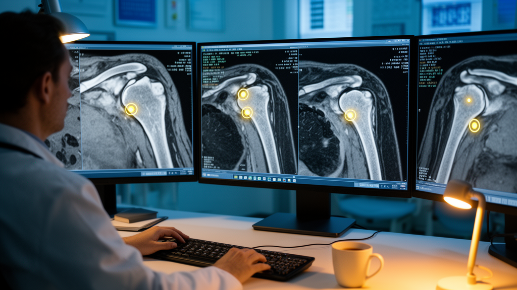
Researchers in Beijing have built a deep-learning network that automatically locates three-dimensional bony landmarks on shoulder CT scans, a step toward automating the mirror-based planning surgeons use before total shoulder arthroplasty. In that planning approach, the healthy, intact shoulder on the opposite side serves as a template for reconstructing the damaged joint, and the technique depends on precisely marking anatomical points such as the humeral head rotation center and the centers of the medullary canal. Doing this by hand is slow and subject to variability both between observers and within the same observer over time, which motivated the team at Beijing Jishuitan Hospital, together with a collaborator from Beijing Estun Medical Technology, to develop an automated alternative.
The study, published in Frontiers in Bioengineering and Biotechnology, was a single-center retrospective analysis of 179 patients whose contralateral shoulder CT scans showed no anatomical damage. The scans were divided at the patient level into a training set of 125 cases, a validation set of 18, and a held-out test set of 36. The architecture, called a dual-decoder 3D BiFormer-UNet, performs two tasks at once: segmenting the humerus and scapula in three dimensions, and localizing 14 anatomical landmarks through heatmap-based regression. A cross-task feature-fusion module passes bone-structural information from the segmentation branch over to the landmark branch, so that the network’s knowledge of where bone structures lie guides its placement of the anatomical points.
Training relied on a composite loss function combining four components: hard-negative-mining heatmap regression, which emphasizes difficult cases during learning; a geometric structure constraint; distance-aware similarity learning; and an auxiliary segmentation loss. The model was trained end-to-end, meaning all components were optimized jointly rather than in separate stages. Performance was measured with the mean radial error (MRE) and the success detection rate (SDR) at thresholds of 2.0, 2.5, 3.0, and 4.0 millimeters, while segmentation quality was assessed with the Dice coefficient. The researchers compared per-case mean radial error against four 3D baseline models using paired statistical tests with Holm–Bonferroni correction for multiple comparisons.
On the 36-case test set, the proposed model achieved a mean radial error of 2.1 ± 1.2 millimeters, with a 95 percent confidence interval of 1.7 to 2.5 millimeters. Its success detection rates were 72.0 percent at the strictest 2.0-millimeter threshold, 81.8 percent at 2.5 millimeters, 90.3 percent at 3.0 millimeters, and 96.7 percent at 4.0 millimeters. The model’s mean radial error was significantly lower than that of 3D U-Net, V-Net, a 3D HRNet adaptation, and H3DE-Net, with all comparisons reaching Holm–Bonferroni-adjusted P values below 0.05. The auxiliary segmentation branch, evaluated on the same test set, reached mean Dice coefficients of 0.83 ± 0.05 for the humerus and 0.78 ± 0.07 for the scapula.
Not all landmarks were localized equally well. Points defined inside the bone rather than on its surface, particularly the humeral head rotation center, the anatomical neck center, and the proximal and distal medullary canal centers, showed larger errors of 2.3 to 2.6 millimeters than most surface landmarks.
The authors conclude that the multi-task framework demonstrated promising landmark localization performance on intact contralateral shoulder CT by combining global anatomical context with bone-structural guidance from the segmentation task. They position the tool as a potential upstream localization component in mirror-based shoulder arthroplasty planning pipelines, where automated landmark detection could replace manual annotation and reduce observer variability.
Several limitations frame these results. The study was retrospective and single-center, and all evaluation was performed on the internal 36-case test set drawn from the same patient population as the training data. The authors themselves state that external validation is required before any clinical use, meaning the reported accuracy figures have not yet been tested on scans from other hospitals, scanners, or patient demographics. The network was also designed and evaluated specifically for shoulders with an intact contralateral side, the population relevant to mirror-based planning, and its behavior outside that setting was not assessed.
Whether that accuracy level is sufficient for clinical planning workflows, and how the system behaves on external data, remain questions for future work rather than findings of the present study.
Subject of Research: Automated three-dimensional detection of shoulder bony landmarks on contralateral CT using a multi-task deep-learning network for arthroplasty planning
Article Title: A multi-task learning-based network for detecting three-dimensional osteometric landmarks of the shoulder joint
Article References: Han, W., Li, F., Ma, C., Zhang, T., Pan, J., & Chen, X. (2026). A multi-task learning-based network for detecting three-dimensional osteometric landmarks of the shoulder joint. Frontiers in Bioengineering and Biotechnology, 14, Article 1948997. https://doi.org/10.3389/fbioe.2026.1948997
Image Credits: AI Generated
DOI: 10.3389/fbioe.2026.1948997
Keywords: deep learning, landmark detection, shoulder arthroplasty, computed tomography, multi-task learning, surgical planning, humerus, scapula, image segmentation, orthopedics, multi-task, learning-based
News Source: Blake Davidson. (October 28, 2026). Deep-learning network pinpoints shoulder landmarks on CT scans. Scienmag.