A supervised deep learning model developed for digital histopathology successfully detected all lymph node metastases from bladder cancer cases while significantly reducing review times for experienced pathologists, according to a validation study at Sahlgrenska University Hospital. In a controlled test evaluating digitized lymph node sections, the artificial intelligence algorithm achieved a sensitivity of 100 percent and a negative predictive value of 100 percent, correctly identifying every whole slide image harboring metastatic disease. Concurrently, deploying model-generated visual overlays during slide review reduced median evaluation times from 9 seconds to 4.2 seconds for the first pathologist and from 12 seconds to 4 seconds for the second pathologist. Both reductions were statistically significant under non-parametric paired testing, demonstrating that an in-house model trained on a constrained institutional dataset can accelerate microscopic assessment without compromising diagnostic sensitivity.
Bladder cancer represents the ninth most frequently diagnosed cancer type worldwide, displaying elevated incidence rates among male populations and within Western regions. Global epidemiological records from 2022 documented approximately 614,000 newly confirmed cases alongside 220,000 associated deaths, positioning the condition as the thirteenth leading cause of mortality across all cancer types. Although bladder tumors encompass diverse benign and malignant histological presentations, the overwhelming majority belong to the urothelial carcinoma category. These neoplasms frequently exhibit distinct morphological subtypes as well as specialized differentiations directed toward squamous or glandular lineages, whereas purely non-urothelial bladder malignancies, such as adenocarcinoma and primary squamous cell carcinoma, remain considerably less common. The clinical management of muscle-invasive bladder cancer routinely centers on radical cystectomy combined with neoadjuvant therapy, accompanied by regional lymph node dissection to evaluate metastatic spread.
Microscopic evaluation of excised regional lymph nodes provides essential prognostic data because metastatic nodal involvement correlates with markedly reduced survival rates following radical cystectomy. Nevertheless, thorough histological examination of dissected nodes demands extensive manual scanning under high magnification, creating an intensive diagnostic workload and contributing substantially to turnaround times. The clinical adoption of whole slide imaging has enabled automated computational pathology tools to assist in primary tumor recognition, histological classification, and molecular alteration prediction across multiple cancer types. Prior investigations established that artificial intelligence systems could uncover subtle nodal micrometastases and match or exceed pathologist performance in breast and lung cancer diagnostics. While a prior multicenter bladder cancer study validated deep learning feasibility across 8,177 whole slide images from over 20,000 lymph nodes, smaller departments frequently lack the digital infrastructure, case volumes, and annotation resources necessary for such expansive frameworks, prompting exploration into localized institutional models.
To determine whether a focused, institution-specific model could provide tangible efficiency gains, investigators gathered archival tissue material from patients who underwent radical cystectomy with lymph node dissection between 2015 and 2024 at Sahlgrenska University Hospital. Patient inclusion required the availability of a representative hematoxylin-eosin stained glass slide derived from formalin-fixed paraffin-embedded tissue. The researchers incorporated all identified patients with verified lymph node metastases, encompassing pathological nodal stages pN1 through pN3, alongside a randomly selected comparative cohort of patients confirmed to have node-negative pN0 disease. Pathological exclusions were applied whenever the primary tumor did not represent urothelial carcinoma, squamous cell carcinoma, or adenocarcinoma, or when metastatic lymph node morphology failed to match the primary tumor. Diagnostic ground truth was established from original pathology reports, with an experienced uropathologist independently re-evaluating every glass slide selected for digitization.
From the eligible clinical cohort, investigators collected 102 hematoxylin-eosin stained glass slides, comprising 71 specimens with confirmed nodal metastases and 31 specimens devoid of metastatic involvement, utilizing a single representative slide per patient case. Each glass specimen was anonymized and digitized in 40x scanning mode at a resolution of 0.23 micrometers per pixel using a 20x objective lens on a Hamamatsu Nanozoomer S210 scanner. After scanning, two whole slide images were excluded due to poor quality, leaving an analytical cohort of 100 slides. The resulting digital files were uploaded directly to the cloud-based Aiforia Create platform, version 6.9, developed by Aiforia Technologies in Helsinki, Finland. Within this digital environment, researchers constructed a supervised deep learning algorithm de novo without prior pretraining weights, configuring convolutional neural network architectures specifically for pixel-wise semantic segmentation tasks.
The technical architecture of the deep learning model relied on two distinct, hierarchically arranged computational layers designed to classify individual image pixels into foreground or background designations. The initial tissue layer was programmed to differentiate biological tissue structures from non-tissue elements, including background glass slide areas, histological preparation artifacts, and cellular debris. The subsequent metastasis layer was trained specifically to separate malignant metastatic tumor cells from normal lymphoid and stromal components situated within lymph nodes. For model development, 66 percent of the total slide collection—consisting of 48 metastatic whole slide images and 18 non-metastatic slides—was allocated to training. Model training was conducted through supervised pixel-level annotations performed exclusively by an expert pathologist across 3,437 designated regions. This progression spanned nine successive training sessions, with intermediate performance metrics analyzed after each session to guide the manual addition of targeted annotations that refined algorithmic boundaries.
Prior to formal testing on whole slide images, the segmentation capabilities of the algorithm were tested against a separate validation dataset of 50 manually delineated regions. This validation set encompassed 16 tissue regions containing confirmed metastatic carcinoma, 32 tissue regions comprising diverse normal nodal structures, and two regions displaying mixed metastatic and normal histological elements. Evaluated against the first independent specialist pathologist validator, the deep learning model achieved a sensitivity of 99.85 percent, a precision or positive predictive value of 99.78 percent, and an F1 score of 99.81 percent. When compared against the second specialist validator, the model recorded an identical sensitivity of 99.85 percent, a precision of 99.49 percent, and an F1 score of 99.67 percent. Crucially, the system accurately detected all validation zones containing metastatic cells as defined by both validators while producing zero false-positive segmentations within purely normal nodal tissue compartments.
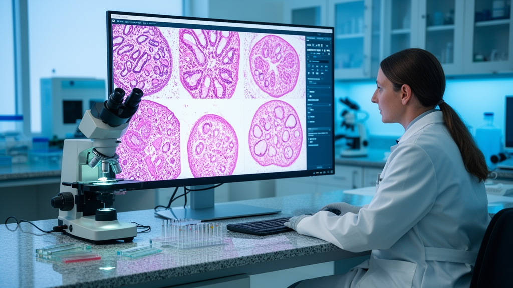
Definitive performance testing took place on an independent test cohort comprising 29 unannotated whole slide images, containing 19 slides with nodal metastases and 10 negative slides without metastatic disease. Two specialist pathologists, each possessing over ten years of diagnostic pathology experience, reviewed every test slide across two distinct experimental rounds. In the initial baseline round, both pathologists evaluated the digitized slides unassisted, using a handheld stopwatch to record assessment times from slide opening to establishing a diagnostic conclusion. Following a two-week washout period designed to minimize visual recall, both clinicians re-evaluated the identical test slides with deep learning assistance, which displayed automated color-coded segmentation masks upon opening. Slide presentation sequences were randomized before each assessment round, and both pathologists remained fully blinded to original diagnostic reports and to each other’s interpretations throughout the entire investigation.
When evaluated against pathological ground truth, the autonomous deep learning model demonstrated a slide-level diagnostic sensitivity of 100 percent, with a 95 percent confidence interval from 82.4 percent to 100 percent. The system simultaneously yielded a negative predictive value of 100 percent, supported by a 95 percent confidence interval of 54.1 percent to 100 percent, successfully flagging all 19 metastatic specimens. However, the model recorded a slide-level specificity of 60 percent, with a 95 percent confidence interval between 26.2 percent and 87.8 percent, alongside a positive predictive value of 82.6 percent, an accuracy of 86.2 percent, and an F1 score of 90.5 percent. Interobserver diagnostic agreement between the two human pathologists was perfect, reflected by a Cohen’s kappa coefficient of 1.00, while interrater concordance between the pathologists and the deep learning system proved substantial, demonstrated by a Krippendorff’s alpha metric of 0.78.
Diagnostic assistance from the deep learning overlay generated marked reductions in slide interpretation duration for both participating specialists across the test cohort. For the first pathologist, median assessment time dropped significantly from 9 seconds down to 4.2 seconds, with a sign test Z score of negative 4.73 and a Wilcoxon signed-rank test Z score of negative 4.44, both yielding p-values below 0.001. Mean review time fell by 70.5 percent, from 24 seconds to 7 seconds overall, decreasing by 73.6 percent for non-metastatic slides and by 62.8 percent for metastatic slides. For the second pathologist, median examination time dropped from 12 seconds to 4 seconds, yielding a sign test Z score of negative 5.00 and a Wilcoxon Z score of negative 4.55, both with p-values below 0.001. The second specialist’s mean assessment time decreased by 87.5 percent, falling from 43 seconds to 5.4 seconds.
The reduction in slide-level specificity in the autonomous model stemmed from small false-positive annotations across four of the ten negative whole slide images. These misidentifications corresponded to benign histological entities, including germinal centers, histiocytes, macrophages, high endothelial venules, and blood vessels, occupying between 0.0025 percent and 0.026 percent of total tissue area with a mean false-positive area of 0.0121 percent. Even on these challenging negative slides, artificial intelligence assistance shortened mean review times by 61.2 percent for the first pathologist and by 89.2 percent for the second pathologist. Study authors noted that with perfect sensitivity, the system is well suited as a triage tool to flag suspicious slides. Limitations include the absence of slides containing solely isolated micrometastases—though the model detected metastatic areas well below 0.01 square millimeters—the lack of external multi-scanner validation, and the need for future studies to assess when cumulative review time savings offset initial training investments.
Subject of Research: Deep learning assistance for histopathological detection of lymph node metastases in bladder cancer
Article Title: Deep learning-assisted detection of lymph node metastases in bladder cancer
Article References: Gorczynski, A., Tarján, B., & Neittaanmäki, N. (2026). Deep learning-assisted detection of lymph node metastases in bladder cancer. Diagnostic Pathology, 21(1), Article 58. https://doi.org/10.1186/s13000-026-01821-8
Image Credits: AI Generated
DOI: 10.1186/s13000-026-01821-8
Keywords: bladder cancer, lymph node metastases, deep learning, histopathology, whole slide imaging, computational pathology, radical cystectomy, urothelial carcinoma, convolutional neural networks, digital pathology, diagnostic sensitivity, pathologist review time, semantic segmentation, Sahlgrenska University Hospital, Aiforia Create, interobserver agreement, false positive foci, triage screening
News Source: Nathaniel Bowman. (November 11, 2026). Deep Learning Model Speeds Bladder Cancer Lymph Node Evaluation Without Missing Metastases. Scienmag.