A deep learning model developed using the YOLOv8 algorithm has successfully detected simulated tooth ankylosis on cone-beam computed tomography scans, achieving an overall precision of eighty percent and a recall of ninety point nine percent. Conducted using laboratory mandibular phantoms, the investigation evaluated whether single-stage deep neural network detectors could automate the identification and boundary delineation of ankylosed root regions on three-dimensional dental radiographs. Tooth ankylosis represents an abnormal fusion of root cementum or superficial dentin directly with the adjacent alveolar bone following the obliteration and replacement of the normal periodontal ligament space by proliferating bone tissue. In physiological conditions, the periodontal ligament consists of specialized connective tissue, collagen fibers, and vascular networks that enable natural tooth mobility and orthodontic displacement. When fusion occurs, physiological movement ceases entirely, potentially impairing eruption paths, causing infra-occlusion of the affected tooth, prompting supra-eruption of opposing dentition, and disrupting adjacent dental alignment.
Clinical diagnosis of ankylosis remains notoriously challenging because routine physical examination cannot directly visualize intra-bony structures, and lack of tooth movement during force application can arise from various etiologies. Conventional two-dimensional radiography, including standard periapical and panoramic projections, frequently fails to reveal localized ankylotic bridges because buccal, palatal, and lingual root surfaces suffer from severe structural superimposition. While three-dimensional cone-beam computed tomography provides multiplanar axial, coronal, and sagittal cross-sections that eliminate adjacent anatomical overlap, manually scrutinizing entire three-dimensional volumes slice by slice is exceptionally laborious and technically demanding even for experienced oral and maxillofacial radiologists. Furthermore, small ankylotic zones often remain undetected when partial volume-averaging artifacts blur minute anatomical features smaller than the acquisition voxel size, while hyperdense lamina dura structures can produce blooming artifacts that generate misleading false-positive interpretations.
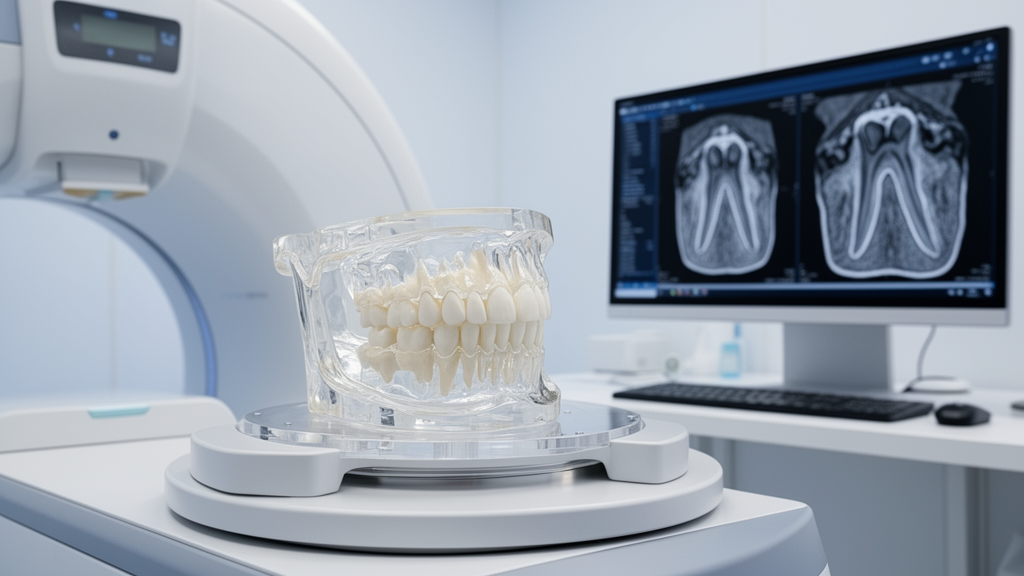
To rigorously investigate an automated detection workflow under standardized conditions, the research team constructed custom mandibular phantoms incorporating sixty extracted human single-rooted teeth, specifically mandibular canines and premolars that had been extracted for reasons unrelated to the investigation. The extracted teeth were thoroughly prepared by immersion in five point twenty-five percent sodium hypochlorite for thirty minutes, mechanically cleaned with brushes to clear residual soft-tissue attachments, and maintained in physiological saline. To replicate the human periodontal ligament space, each tooth was mounted individually in type one dental stone for mechanical stability and uniformly coated with a one-millimeter thermoplastic sheet using an automated vacuum former. Experimental measurements confirmed that this protocol achieved an average simulated ligament layer thickness of one hundred twenty-five plus or minus twenty-five micrometers across the coronal, middle, and apical thirds of the roots, closely matching natural anatomical dimensions.
Simulated ankylotic defects were subsequently created on all sixty specimens by carefully excising a precise two-by-two millimeter square of the thermoplastic coating using a number fifteen-C surgical scalpel. These defects were randomly distributed across cervical, middle, or apical root segments situated on buccal, lingual, mesial, or distal surfaces. To establish a realistic bony architecture, silicone impressions of a three-dimensional printed human mandible were cast using a specialized slurry formulation combining type three dental stone and ground rice mixed in a two-to-one ratio, effectively mimicking human trabecular bone patterns. The coated teeth were mounted into ten mandibular models, with six teeth randomly assigned to each model. To ensure seamless continuity and intimate contact without air voids, an additional thin stone-rice mixture was carefully painted directly onto the exposed root defect windows before final casting and setting.
Radiographic acquisition was performed by submerging the completed phantoms in a water-filled cylindrical polypropylene container measuring sixteen by sixteen by seventeen point seven centimeters to simulate surrounding soft tissue attenuation, maintaining a water layer between twenty and fifty millimeters around each specimen. Imaging data were collected across two distinct scanner platforms to support independent evaluation. The primary training dataset comprised forty-two teeth across seven imaging volumes, yielding three thousand eight hundred twenty-two axial sections captured on a NewTom VGi Evo scanner configured with an eight-by-eight centimeter field of view, zero point one hundred fifty millimeter voxel size, one hundred ten kilovolts peak, eleven point eighty-six milliamperes, and a five point four second exposure time. The secondary test dataset comprised eighteen teeth across three volumes, totaling one thousand one hundred forty-nine axial sections obtained on a Carestream CS 8100SC 3D unit operating with an eight-by-nine centimeter field of view, zero point one hundred fifty millimeter voxel size, ninety kilovolts peak, four milliamperes, and a fifteen second exposure time.
Ground-truth segmentation masks were manually delineated across every sequential axial slice of each volumetric scan by a senior oral and maxillofacial radiology resident and verified by an experienced radiologist using ITK-SNAP software. The interactive three-dimensional brush tool was intentionally disabled during annotation to prevent false-positive over-segmentation, relying instead on small manual paintbrush sizes adjusted between three and five pixels to capture minute boundary interfaces. Axial delineations were systematically cross-examined and confirmed against coronal and sagittal reconstructions. For deep learning development, the authors deployed the Ultralytics YOLOv8 architecture, a single-stage convolutional neural network comprising backbone, neck, and detection head components. Evaluating network complexity, latency, and predictive performance across nano, small, medium, large, and extra-large configurations, the researchers selected the YOLOv8 medium model as the optimal baseline balancing parameter scale, inference speed, and localization precision.
Volumetric data were preprocessed by normalizing radiodensity values within an intensity window spanning six hundred to sixteen hundred Hounsfield units. Model generalization was supported through data augmentation procedures incorporating horizontal flips, random contrast adjustments, and spatial rotations. Network training was executed in Python using an NVIDIA 3060Ti graphics processing unit, governed by the Adam optimization algorithm with a batch size of sixteen, an initial learning rate of zero point zero zero one, and an early stopping threshold set at one hundred epochs. Training loss was continually supervised using batch loss calculations and adaptive learning rate adjustments, successfully descending below ten percent after one hundred training cycles. Cross-validation was managed via a fivefold internal validation framework within the training cohort to preserve maximal training data density without sacrificing hyperparameter stability.
Quantitative assessment on the independent test dataset demonstrated robust diagnostic and localization capability across all monitored evaluation metrics. The trained YOLOv8 model achieved a precision of eighty percent, confirming its capacity to identify true ankylotic bridges while minimizing false-positive detections. The model yielded a recall of ninety point nine percent, reflecting high sensitivity in capturing true lesions across complex root geometries, alongside an overall F1 score of eighty-three point nine percent. Spatial overlap evaluation produced a Dice similarity coefficient of eighty-two percent and an intersection over union of seventy-four percent between automated predictions and reference annotations. Comparative visual analysis of contoured outputs demonstrated high spatial alignment between model segmentations and radiologist ground truth, confirming reliable localization across sequential axial slices.
These experimental outcomes demonstrate the clinical promise of deploying single-stage object detection architectures for automated screening in dental radiology. Unlike conventional classification models such as ResNet architectures, which only indicate the global presence or absence of pathology within a full image slice without locating boundaries, detection networks explicitly output bounding coordinates and spatial masks. This precise localization is critical for orthodontists and oral surgeons who must determine the exact anatomical position and vertical extent of fusion before initiating tooth movement or surgical luxation. Furthermore, because YOLOv8 requires substantial computational power primarily during training phases, trained models can easily execute real-time inference on standard desktop computers routinely found in private dental offices and outpatient diagnostic centers.
Despite these encouraging findings, several methodological limitations must be considered when interpreting the study results. The investigation relied exclusively on single-rooted mandibular canines and premolars embedded in synthetic phantom matrices, excluding multi-rooted molars where complex furcation anatomies present significant diagnostic hurdles. Moreover, the phantom environment could not reproduce clinical sources of artifacts, such as patient motion during scanning, which may affect image quality and detection performance. The researchers recommended that future studies evaluate multi-rooted and impacted teeth, assess models combining in vitro and in vivo datasets, and directly compare algorithmic performance against radiologists.
Subject of Research: Deep learning detection of tooth ankylosis using YOLOv8 on cone-beam computed tomography phantom scans
Article Title: A phantom study on the evaluation of the YOLOv8 deep learning model in the detection of tooth ankylosis on cone-beam computed tomography images
Article References: Moshfeghi, M., Shayesteh, S. P., Valizadeh, S., Pakravan, P., & Nikmanesh, N. (2026). A phantom study on the evaluation of the YOLOv8 deep learning model in the detection of tooth ankylosis on cone-beam computed tomography images. Head & Face Medicine, 22(1), Article 55. https://doi.org/10.1186/s13005-026-00607-z
Image Credits: AI Generated
DOI: 10.1186/s13005-026-00607-z
Keywords: Tooth ankylosis, YOLOv8, Deep learning, Cone-beam computed tomography, CBCT, Artificial intelligence, Dental radiology, Periodontal ligament, Object detection, Image segmentation, Phantom study, Precision and recall, Dice coefficient, Medical image analysis, Convolutional neural network, Oral and maxillofacial radiology
News Source: Blake Davidson. (November 20, 2026). Deep Learning Model Detects Simulated Tooth Ankylosis in Phantom CBCT Study. Scienmag.