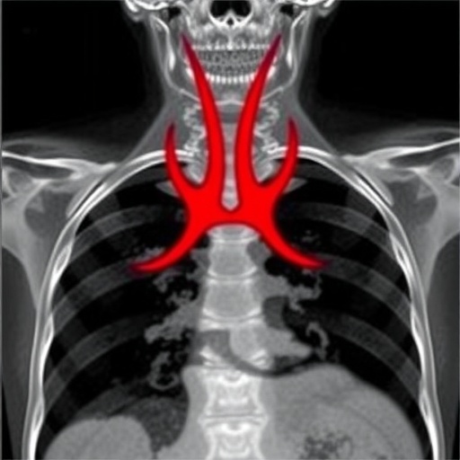
In a groundbreaking case report that pushes the boundaries of forensic and clinical medicine, researchers have documented a fatal incident revealing the intricate and often underestimated dangers of bronchoscopy procedures. The study centers on a rare and devastating complication: an obstructive fibrinous tracheal pseudomembrane, which developed secondary to a hemorrhagic rupture of the trachea during a bronchoscopy. This report, unparalleled in its detail and multimodal imaging approach, sheds new light on the critical vulnerabilities of the tracheal structure during invasive respiratory diagnostics and therapeutic interventions.
The bronchoscopy, a commonly employed endoscopic technique used to visualize the inside of the airways, has been a mainstay diagnostic tool, especially for pulmonary pathologies. However, this case underscores the potential life-threatening risks intrinsic to the procedure, which are not fully appreciated outside specialized medical circles. The tracheal rupture in this patient’s case triggered a pathological cascade that culminated in the formation of an obstructive fibrinous pseudomembrane, effectively sealing the airway and leading to rapid clinical deterioration and death.
The term “fibrinous pseudomembrane” refers to a dense, mesh-like accumulation of fibrin—a protein that plays a key role in blood clotting and tissue repair—along with inflammatory cells, that adheres tightly to the mucosal surfaces of the trachea. This pathological entity can obstruct airflow significantly, converting what might have been a survivable injury into a fatal airway emergency. The pseudomembrane’s development in this context represents a formidable physiological response to trauma but with catastrophic consequences in a confined anatomical space.
Employing a sophisticated suite of post-mortem imaging techniques, including high-resolution computed tomography (CT) scans and possibly magnetic resonance imaging (MRI), the researchers were able to delineate the exact anatomical disruptions and pathological sequelae. The multimodal imaging approach facilitated an unprecedented visualization of the intratracheal pathology, illustrating the extent of the hemorrhagic rupture and the pseudomembrane architecture. This methodological framework not only enhances accuracy in forensic evaluations but also facilitates a deeper understanding of airway pathology mechanisms in clinical practice.
The hemorrhagic tracheal rupture, identified as the primary event during the bronchoscopy, is an exceedingly rare but catastrophic procedural complication. Such ruptures usually stem from mechanical trauma inflicted by the bronchoscope or associated instrumentation, especially in patients with pre-existing tracheal frailty or other risk factors like chronic inflammation or prior radiotherapy. The hemorrhage associated with these ruptures exacerbates tissue injury and promotes an aggressive inflammatory response, fostering fibrin deposition and pseudomembrane genesis.
Clinicians and forensic experts alike will find this case vital for appreciating the nuanced interplay between mechanical injury and subsequent pathological responses within the respiratory tract. It explains, in stark terms, how even meticulously conducted bronchoscopies bear inherent risks that can spiral into fatal outcomes under rare but critical circumstances. The case acts as a wake-up call to enhance procedural vigilance and patient monitoring during bronchoscopic interventions.
Moreover, the case exemplifies the paradigm shift in forensic pathology towards integrating advanced imaging techniques with traditional autopsy methods. The non-invasive imaging not only provides supplemental verification of autopsy findings but also preserves crucial diagnostic information that can guide clinical improvements and safety protocols. In this case, imaging was instrumental in correlating the macroscopic findings with histopathological evidence, offering a comprehensive pathological narrative.
The documentation meticulously traces the patient’s clinical trajectory, illustrating how the initial procedure-induced trauma went unrecognized, allowing the fibrinous pseudomembrane to evolve unchecked. This temporal dimension to the case emphasizes the critical need for post-procedural surveillance strategies to detect early signs of airway compromise. Clinicians should be alert to sudden respiratory distress following bronchoscopy and consider advanced imaging diagnostics promptly.
From a scientific standpoint, this case contributes significantly to the literature by elucidating the pathophysiological processes underlying pseudomembrane formation in the airways. The researchers propose a model where fibrin deposition is not simply a response to bleeding but an active pathological process magnified by local tissue hypoxia and sustained inflammation. These insights open avenues for targeted therapeutic interventions aiming to mitigate such exuberant fibrinous reactions following airway injury.
The fatal outcome detailed in this report also sparks ethical and medico-legal discussions regarding informed consent, procedural risk disclosure, and the thresholds for intervention. It compels healthcare providers to balance diagnostic necessity against potential procedural hazards, particularly in vulnerable populations. The forensic implications extend into medico-legal investigations where cause-of-death determination must consider iatrogenic factors and procedural complications meticulously.
In terms of procedural implications, the necessity for gentle bronchoscope manipulation and real-time monitoring is re-emphasized. The current case sets a precedent for developing guidelines to recognize early signs of airway rupture and obstruction during the procedure. Incorporating real-time imaging or novel sensor technologies might serve as preventive innovations to minimize mechanical trauma during bronchoscopies.
The reliance on advanced imaging techniques in this case may also influence future training for pulmonologists, surgeons, and forensic pathologists, integrating radiological expertise into routine procedural and post-procedural practice. The synergy between clinical medicine and imaging is poised to improve patient safety and diagnostic precision, which could, in time, reduce morbidity and mortality associated with invasive airway procedures.
In conclusion, this fatal case report offers the medical and forensic communities an instructive exemplar of rare but recognized risks associated with bronchoscopy. The fusion of clinical insight, pathological analysis, and cutting-edge imaging forged a comprehensive understanding of the sequence of events leading to death. Such knowledge is invaluable for shaping safer clinical protocols, enhancing diagnostic acumen, and refining forensic methodologies.
The implications of this research extend far beyond a single case, signaling a shift towards heightened awareness, improved procedural caution, and interdisciplinary collaboration. It stands as a stark reminder that in the quest for diagnosis and intervention, the complexity of human anatomy and pathophysiologic responses demands both precision and prudence.
As the field advances, the insights garnered here will hopefully catalyze preventative strategies and technological innovations to avert similar tragic outcomes. This multimodal imaging case report may well become a cornerstone reference in the domains of legal medicine, pulmonary diagnostics, and interventional safety standards.
Subject of Research: Fatal airway obstruction due to fibrinous tracheal pseudomembrane formation following hemorrhagic tracheal rupture induced by bronchoscopy.
Article Title: Fatal case of obstructive fibrinous tracheal pseudomembrane caused by hemorrhagic tracheal rupture during bronchoscopy: a multimodal post-mortem imaging case report.
Article References:
Zhao, X., Chen, Z., Wang, B. et al. Fatal case of obstructive fibrinous tracheal pseudomembrane caused by hemorrhagic tracheal rupture during bronchoscopy: a multimodal post-mortem imaging case report. Int J Legal Med (2025). https://doi.org/10.1007/s00414-025-03617-9
Image Credits: AI Generated
Tags: airway obstruction due to pseudomembranebronchoscopy procedure risksclinical implications of tracheal injuriesfatal bronchoscopy case reportforensic medicine advancementshemorrhagic tracheal ruptureinvasive respiratory diagnostics dangersobstructive fibrinous tracheal pseudomembranepost-mortem imaging in forensicsrespiratory therapeutic interventions riskstracheal rupture complicationstracheal structure vulnerabilities
