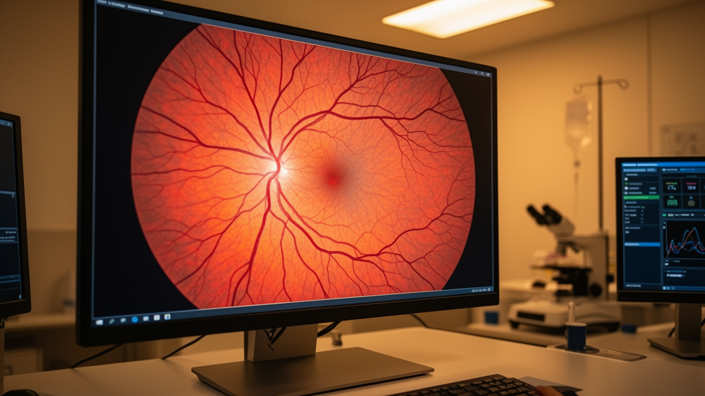
A research team from the Vellore Institute of Technology has developed a multistage image processing framework designed to enhance retinal fundus photographs while maintaining high structural similarity. The investigators established the sequential framework to improve the visibility of delicate retinal structures and to facilitate automated ophthalmic image analysis across diverse image-quality conditions.
The proposed D-CGRet framework operates through a hybrid, multistage enhancement pipeline engineered to systematically counter noise degradation, local contrast limitations, and non-uniform illumination. To achieve these corrections without sacrificing critical retinal features, the sequential processing workflow combines filter-based denoising techniques, Contrast Limited Adaptive Histogram Equalization, Adaptive Gamma Correction with Weighting Distribution, and Multi-Scale Retinex illumination enhancement. Each computational stage in the sequential pipeline is organized to address distinct image-quality deficiencies while actively preserving the relevant retinal information required for downstream analysis.
In the initial phase of experimental evaluation, the researchers examined the denoising capabilities of the framework across 45 noise-augmented test images. This first experiment specifically investigated the pipeline’s operational performance under Gaussian noise, salt-and-pepper noise, and speckle noise conditions. Within this denoising stage, four distinct digital filters were independently evaluated: median filters, Gaussian filters, Wiener filters, and average filters. Following the noise suppression stage, the framework carried out Contrast Limited Adaptive Histogram Equalization directly on the Value component within the hue, saturation, and value color space, followed sequentially by adaptive gamma correction and Multi-Scale Retinex-based illumination enhancement.
To further assess operational performance beyond synthetic noise conditions, additional experiments examined selected original retinal fundus photographs drawn from two established benchmark collections: the Fundus Photo Enhancement dataset and the Digital Retinal Images for Vessel Extraction dataset. The selected image sets permitted the authors to evaluate the framework across four distinct image-quality conditions, namely poor-quality fundus images, normal images, over-contrasted photographs, and dark retinal images. These varying visual baselines allowed the research team to systematically observe how effectively the enhancement pipeline responded to differing forms of illumination and contrast limitations.
The performance of the D-CGRet framework was evaluated through a multi-angle assessment strategy that combined comparative analyses against existing enhancement techniques, direct visual inspection of the enhanced outputs, and specialized retinal blood-vessel analysis comparing the original images with the processed outputs. To ensure a thorough quantitative evaluation across the processed images, the investigators recorded six distinct performance metrics: Peak Signal-to-Noise Ratio, Structural Similarity Index Measure, Correlation Coefficient, Edge Preservation Index, Contrast Improvement Index, and entropy across the test datasets.
The quantitative findings confirmed that the proposed framework achieved high structural fidelity while carrying out illumination and contrast adjustments. Achieving Structural Similarity Index Measure values of 0.94 in normal fundus images and 0.92 in dark fundus images, the D-CGRet system demonstrated high structural similarity under varied image-quality conditions. The researchers noted that these combined experimental contributions demonstrate a systematic evaluation of noise suppression, contrast enhancement, illumination correction, and the preservation of vascular details necessary for ophthalmic evaluation.
Although the experimental results demonstrate the potential of D-CGRet to improve retinal fundus images for the specific cases studied, the authors emphasized critical limitations regarding the study sample and overall generalizability. Consequently, Jaiswar and Gupta concluded that the experimental findings need to be further validated on larger and more diverse datasets before the framework can be generalized across wider automated ophthalmic workflows.
Subject of Research: Retinal fundus image enhancement and denoising
Article Title: D-CGRet: a multistage hybrid framework for denoising and contrast improvement in the retinal fundus image
Article References: Jaiswar, S., & Gupta, S. (2026). D-CGRet: a multistage hybrid framework for denoising and contrast improvement in the retinal fundus image. Frontiers in Ophthalmology, 6, Article 1946223. https://doi.org/10.3389/fopht.2026.1946223
Image Credits: AI Generated
DOI: 10.3389/fopht.2026.1946223
Keywords: retinal fundus imaging, image enhancement, D-CGRet, denoising filters, contrast limited adaptive histogram equalization, adaptive gamma correction, multi-scale retinex, ophthalmic image analysis, structural similarity index measure, peak signal-to-noise ratio, edge preservation index, contrast improvement index, DRIVE dataset, Fundus Photo Enhancement dataset, retinal blood-vessel analysis, illumination correction, HSV color space
News Source: Ophelia Keating. (November 18, 2026). D-CGRet Framework Enhances Retinal Images With High Structural Similarity in Tests. Scienmag.