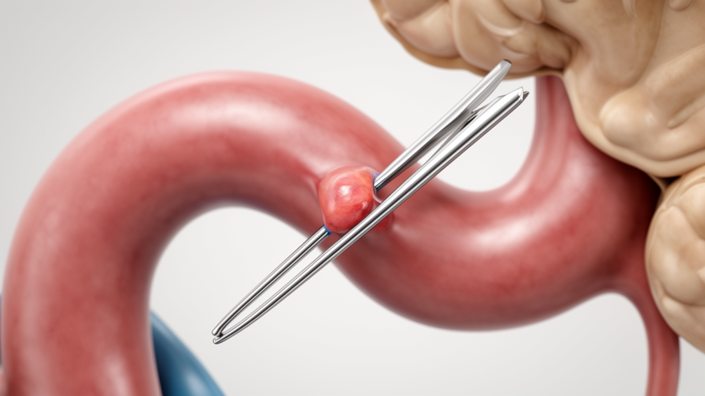
In a correspondence published in the Egyptian Journal of Neurosurgery, researchers addressed methodological and analytical questions regarding the relationship between the anatomical course of the first segment of the middle cerebral artery and the frequency of temporary clipping during aneurysm surgery. Writing in response to commentary by Munir and colleagues, the surgical team, including Daniel Serfaty-Fonseca and Eberval Gadelha Figueiredo, clarified specific evaluation procedures and statistical interpretations stemming from their previously published cohort study. The authors emphasized that the observed anatomical associations serve primarily as hypothesis-generating observations rather than definitive causal mechanisms, calling for careful consideration of vascular geometry alongside individual patient and aneurysm characteristics when planning operative proximal control.
The central comparison in the investigation evaluated whether a curved middle cerebral artery first segment (M1) altered the likelihood of placing a temporary clip compared with a straight vessel configuration. In the analyzed patient cohort, temporary vascular occlusion was documented in 10 out of 39 surgical cases presenting with curved M1 morphology. In contrast, temporary clipping occurred in only 3 out of 47 surgical cases characterized by a straight M1 segment. This distribution yielded an unadjusted cross-product odds ratio of 5.06, indicating an apparent elevation in clipping frequency among individuals with curved vascular anatomy during surgical management.
To assess the precision of this statistical estimate, the authors recalculated the 95 percent confidence interval using the conventional asymptotic log-Wald formulation, reproducing the reported range of 1.28 to 19.96. While binary hypothesis testing was conducted using Fisher’s exact test to accommodate the small sample distribution, the notable width of the resulting confidence interval highlights substantial statistical uncertainty regarding the true magnitude of the association. The investigative team acknowledged that the limited absolute number of clipping events, thirteen instances across eighty-six total patients, appropriately informed their decision to present descriptive, unadjusted statistical analyses rather than adjusted models.
Addressing inquiries regarding the objectivity of morphological classification, the authors clarified that preoperative vascular imaging was reviewed independently by two clinical evaluators who were unaware of temporary-clipping status during classification. The authors noted that this blinded classification protocol was implemented to resolve concerns that knowledge of operative outcomes could influence image interpretation, a detail not explicitly stated in the published Methods. High interobserver agreement between the two evaluators further supported the reproducibility of the anatomical categorization within their patient cohort.
The authors also discussed the clinical nuance distinguishing elective temporary clipping from reactive clipping performed in response to intraoperative aneurysm rupture. Although the original database allows elective clipping for planned proximal control to be distinguished from clipping executed following intraoperative rupture, these distinct indications were not analyzed separately in the published report. Because the primary endpoint described overall temporary-clipping use in aggregate, the authors agreed that examining indications and the chronological sequence of operative events in subsequent studies will help determine how much of the association relates to planned control versus rupture management.
Finally, the correspondence noted that the underlying patient cohort underwent surgical treatment under a single-surgeon design across a multi-year timeframe extending from 2006 through 2018. Complete surgical dates, including exact day, month, and year, were systematically recorded and remain retrievable through study collaborators, although calendar time was not incorporated into the published analysis. The authors observed that while a single-surgeon practice provides procedural consistency, it does not fully account for potential changes in experience, neuroimaging, or operative judgment over time. Assessing temporal distributions and clipping indications in future work will help establish the true clinical significance and generalizability of M1 segment morphology.
Subject of Research: Middle cerebral artery M1 segment morphology and temporary clipping in aneurysm surgery
Article Title: Authors’ response to “M1 segment morphology and temporary clipping in middle cerebral artery aneurysm surgery”
Article References: Serfaty-Fonseca, D., Tzu, W. H., da Silva, S. A., Serfaty, D. D. S. M., de Holanda, L. S., & Figueiredo, E. G. (2026). Authors’ response to “M1 segment morphology and temporary clipping in middle cerebral artery aneurysm surgery”. Egyptian Journal of Neurosurgery, 41(1), Article 199. https://doi.org/10.1186/s41984-026-00707-7
Image Credits: AI Generated
DOI: 10.1186/s41984-026-00707-7
Keywords: middle cerebral artery, M1 segment, aneurysm surgery, temporary clipping, vascular morphology, proximal control, odds ratio, Fisher's exact test, log-Wald method, interobserver agreement, preoperative imaging, intraoperative rupture, elective clipping, single-surgeon study, curved M1, straight M1, Egyptian Journal of Neurosurgery
News Source: Ophelia Keating. (November 19, 2026). Curved M1 Segment Linked to Higher Temporary Clipping Odds in Aneurysm Surgery. Scienmag.