Bringing faster 3D imaging for biomedical researches
Credit: The Chinese University of Hong Kong
The research result has been published in the journal Optics Letters recently.
Activities of neurons are generally completed on a time scale of 10 milliseconds, which makes it hard for conventional microscopes to observe these phenomena directly. This new compressive sensing two-photon microscopy can be applied to 3D imaging of the nerve distribution of living things or to monitoring activities from hundreds of neurons simultaneously.
New multi-focus laser scanning method to break the scanning speed limit of two-photon microscope
Two-photon microscopy works by delivering ultrafast pulses of infrared laser light to the sample, where it interacts with fluorescent labels to create an image. It is extensively used for biological researches because of its ability to produce high-resolution 3D images up to a depth of one millimeter in a living tissue. These advantages, however, come with a limited imaging speed of the two-photon microscopy because of the weak fluorescent signal.
To speed up scanning, the research team developed a multi-focus laser illumination method that uses a digital micromirror device (DMD). The research solves the problem of conventional DMD being unusable to work with ultrafast laser, enabling them to be integrated and used in beam shaping, pulse shaping, and two-photon imaging.
The DMD generates 30 points of focused laser light on randomly selected locations within a specimen. The position and intensity of each point of light are controlled by a binary hologram that is projected onto the device. During each measurement, the DMD reflashes the hologram to change the position of each focus and records the intensity of the two-photon fluorescence with a single-pixel detector. Although, in many ways, the DMD multi-focus scanning is more flexible and faster than traditional mechanical scanning, the speed is still limited by the DMD’s refresh rate.
Combining the compressive sensing algorithm to further improve the imaging speed
The researchers further increased the imaging speed in this research by combining multi-focus scanning with compressive sensing. This approach enables image acquisition with fewer measurements. This is because it carries out image measurement and compression in a single step and then uses an algorithm to rebuild the images from the measurement results. For two-photon microscopy, it can reduce the number of measurements by between 70% and 90%.
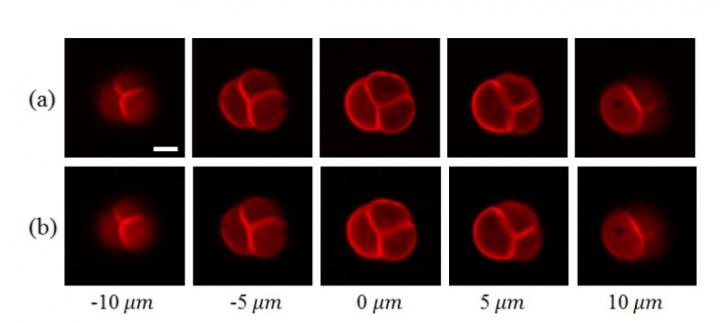
After conducting a simulation experiment to demonstrate the new method’s performance and parameters, the researchers tested it with two-photon imaging experiments. These experiments demonstrated the technique’s ability to produce high-quality 3D images with high imaging speeds from any field of view. For example, they were able to acquire 3D images from a pollen grain, in just 0.55 seconds. The same images acquired with traditional point scanning took 2.2 seconds.
Prof. Shih-Chi Chen said, “This method achieved a three to five times enhancement in imaging speed without sacrificing the resolution. We believe this novel approach will lead to new discoveries in biology and medicine, such as optogenetics. The team is now working to further improve the speed of the reconstruction algorithm and image quality. We also plan to use the DMD together with other advanced imaging techniques, which allows imaging in deeper tissues.”
###
Media Contact
Angela Wan
[email protected]
852-394-33916
Original Source
https:/


