
In the dynamic and ever-evolving field of forensic medicine, precise age estimation remains a cornerstone for both legal and humanitarian applications. Recent advances have underscored the pivotal role of medical imaging, particularly computed tomography (CT), in refining this intricate process. A groundbreaking study has emerged highlighting the significant impact of patient positioning during CT scans of the medial clavicular epiphysis—a critical anatomical marker used for forensic age assessment. The researchers, Kuhnen, Müller, Schmeling, and colleagues, have identified the ‘raised arms position’ as a superior technique, offering enhanced clarity and reliability. This development promises to recalibrate forensic standards globally, streamlining age estimation protocols and bolstering judicial accuracy.
The medial clavicular epiphysis has long been recognized as a reliable skeletal indicator for age estimation, especially in individuals approaching adulthood. Traditional radiographic methods have faced limitations due to overlapping anatomical structures and variable ossification patterns that complicate image interpretation. By harnessing CT technology—a modality revered for its high-resolution cross-sectional imaging—the anatomical intricacies of the medial clavicular epiphysis can be visualized with unprecedented precision. Nevertheless, the positioning of the subject during scanning has often been overlooked, with a default ‘arms-down’ posture commonly utilized. The study’s authors challenge this convention, demonstrating the enhanced efficacy of a ‘raised arms’ posture.
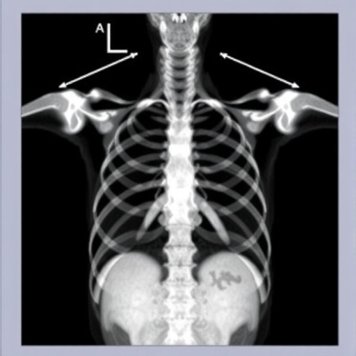
In applying the raised arms position, patients elevate their upper limbs above their head, effectively altering the spatial orientation of the clavicle relative to adjacent structures. This positional adjustment serves to mitigate superimposition artifacts and reduces the anatomical overlap that typically obscures the medial clavicular epiphysis in standard scans. By minimizing structural convergence, the raised arms position facilitates clearer delineation of epiphyseal ossification stages, thereby refining age estimation accuracy. The study’s quantitative analyses corroborate the qualitative improvements observed, suggesting a paradigm shift in forensic imaging protocols.
.adsslot_sXRNnOlV7H{width:728px !important;height:90px !important;}
@media(max-width:1199px){ .adsslot_sXRNnOlV7H{width:468px !important;height:60px !important;}
}
@media(max-width:767px){ .adsslot_sXRNnOlV7H{width:320px !important;height:50px !important;}
}
ADVERTISEMENT
Encapsulating adult skeletal maturation, the medial clavicular epiphysis exhibits distinct radiological features that evolve predictably with chronological age. Ossification initiation, progression, and eventual fusion stages form a timeline against which biological age can be extrapolated. However, the precision of these assessments hinges on the visibility and contrast of the epiphyseal plate and its surrounding cortical bone. The raised arms technique significantly enhances these imaging characteristics, providing forensic experts with a more detailed morphological canvas to inform their evaluations. This has notable implications for cases where legal thresholds hinge on exact age determination.
Forensic age estimation is not merely an academic exercise but a critical tool in numerous legal contexts, including the identification of undocumented minors, criminal responsibility assessments, and the adjudication of asylum claims. The methodologies employed must therefore strike a balance between scientific rigor, reproducibility, and non-invasiveness. CT scans, despite their radiation dose considerations, remain the gold standard in assessing internal skeletal structures non-destructively. Optimizing patient positioning to extract maximal information from each exposure aligns with the ethical imperative to minimize patient risk while maximizing diagnostic yield.
Moreover, the study’s meticulous approach to evaluating patient positioning underscores the broader necessity of standardizing imaging protocols in forensic practice. Variability in scan acquisition parameters can introduce discrepancies that compromise comparability across cases and jurisdictions. By advocating for the raised arms position, the authors provide a clear, actionable recommendation that can enhance consistency and reliability. This standardization effort extends beyond mere technical preference, potentially influencing forensic guideline development and international consensus statements.
The imaging technique itself—a multidetector CT acquisition—provides volumetric data enabling three-dimensional reconstruction and multiplanar reformation of the medial clavicular epiphysis. These advanced imaging capabilities allow forensic radiologists to assess ossification in various planes, circumventing limitations imposed by traditional two-dimensional radiography. Combined with the improved anatomical visualization afforded by the raised arms position, this synergistic methodology elevates forensic age assessment into a new era of precision and clarity. The study provides compelling evidence supporting its routine implementation.
Interestingly, the researchers also investigated the inter-observer reliability of their imaging protocol. Enhanced visibility of the epiphyseal structures in the raised arms scans correlated with improved concordance rates among forensic experts. This statistical affirmation further validates the clinical utility of the positioning adjustment. The reduction in subjective interpretation variability heightens confidence in forensic reports, which in turn bolsters their judicial weight. As forensic age estimation findings increasingly influence decisions that profoundly affect individuals’ lives, such improvements in methodological robustness are invaluable.
Beyond the primary findings, the study touches on radiation dose optimization strategies accompanying the patient positioning recommendation. Elevated arms positioning can decrease the necessity for repeat scans caused by suboptimal visualization, thereby indirectly contributing to dose reduction. Additionally, refined imaging parameters tailored to this posture could further minimize exposure while preserving diagnostic quality. This dual benefit addresses ongoing concerns regarding radiation safety and aligns with ALARA principles (As Low As Reasonably Achievable) fundamental to medical imaging practice.
Clinicians and forensic scientists should also appreciate the practical implications regarding patient comfort and scan workflow. While raising arms may seem a minor adaptation, it necessitates clear patient instructions and possible adjustments in immobilization devices to maintain stability during acquisition. The research team elaborates on manageable protocols facilitating this adjustment without inducing patient discomfort or compromising image integrity. Adoption of such methods requires interdisciplinary cooperation between radiologists, technicians, and forensic experts, emphasizing the collaborative nature of forensic imaging.
As forensic age estimation advances in sophistication, integrating emerging technological innovations such as machine learning and image segmentation algorithms holds promise. The high-quality images obtained through the raised arms CT positioning could serve as superior datasets for training artificial intelligence models designed to automate and enhance ossification stage classification. This technical evolution might democratize forensic expertise, providing accessible, objective tools applicable even in resource-limited settings. The foundational work on optimal imaging positioning thus catalyzes future research and development trajectories.
The broader forensic community is likely to welcome these insights with enthusiasm, particularly given the growing global demand for accurate age assessments amidst complex migration and legal challenges. The raised arms CT protocol represents a relatively simple yet impactful modification that leverages existing technology to yield markedly improved results. As forensic science continually strives to balance innovation with pragmatic application, such contributions exemplify how methodical reevaluation of longstanding practices can generate meaningful progress.
In conclusion, the study spearheaded by Kuhnen and colleagues exemplifies rigorous scientific inquiry with profound practical implications. By demonstrating the superiority of the raised arms position during CT imaging of the medial clavicular epiphysis, they have paved the way for enhanced accuracy, reproducibility, and safety in forensic age estimation. This research not only refines a critical diagnostic tool but also underscores the importance of detailed attention to imaging methodology. Its uptake across forensic institutions promises to elevate the reliability of age determination, ultimately strengthening the integrity of judicial processes worldwide.
As forensic medicine intersects increasingly with human rights and social justice issues, the importance of precise biological age estimation cannot be overstated. Technical advancements such as those presented here embody the potential of scientific innovation to inform and improve real-world outcomes. The integration of optimized patient positioning in CT protocols may appear incremental but holds disproportionate promise in enhancing forensic evidence quality. Future guidelines and training programs are expected to incorporate these findings, marking a significant step forward for practitioners and the individuals whose lives depend on their expertise.
Continued research will undoubtedly explore further refinements, including adaptation across diverse populations and age ranges, comparative efficacy against alternative imaging modalities, and cost-benefit analyses of widespread protocol implementation. Nonetheless, the clarity and robustness of evidence supporting raised arms positioning offer a compelling impetus for immediate consideration and adoption. This exemplifies how attention to seemingly subtle technical details can revolutionize forensic standards and ultimately improve societal trust in scientific methods underpinning the justice system.
Subject of Research: Forensic age estimation using CT imaging of the medial clavicular epiphysis and the impact of patient arm positioning.
Article Title: CT of the medial clavicular epiphysis for forensic age estimation – raised arms position recommended.
Article References:
Kuhnen, S.C., Müller, M., Schmeling, A. et al. CT of the medial clavicular epiphysis for forensic age estimation – raised arms position recommended. Int J Legal Med (2025). https://doi.org/10.1007/s00414-025-03521-2
Image Credits: AI Generated
Tags: advances in forensic medicineage estimation protocols in forensic applicationsanatomical visualization in forensic sciencechallenges in traditional radiographic methodsCT scans for forensic age estimationenhanced clarity in medical imaginghigh-resolution imaging techniquesimproving judicial accuracy through imagingmedial clavicular epiphysis analysismedical imaging in age assessmentraised arms position in CT imagingskeletal indicators for age determination

