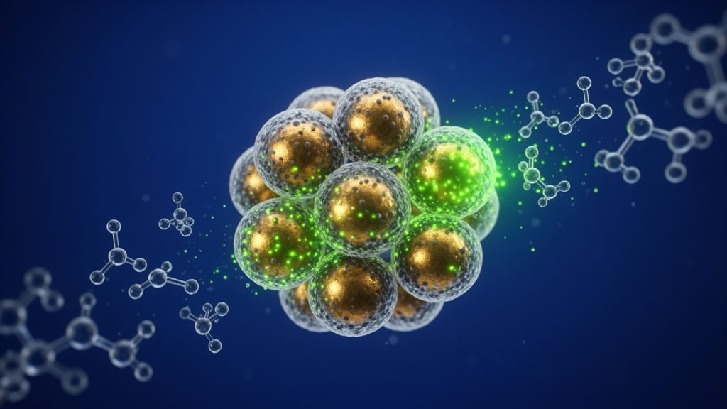
Researchers have designed and synthesized a multi-component Au@SiO2/CQD nanocomposite material that operates as a rapid, sensitive, and stable electrochemical sensing platform for the detection of hydrogen peroxide. Engineered by adsorbing hydrothermally derived carbon quantum dots onto the surface of core-shell gold-silica nanoparticles, the resulting composite integrates the high electrical conductivity and catalytic activity of gold cores, the spacer and surface area benefits of silica shells, and the electrochemiluminescent and electronic attributes of carbon quantum dots. When modified onto the surface of a glassy carbon electrode, this nanostructured material enabled the rapid amperometric determination of hydrogen peroxide in an aqueous electrolyte at concentrations as low as 0.2 millimolar. The modified electrode demonstrated high current sensitivity, rapid signal stabilization achieved within four seconds of analyte introduction, robust cycling performance across repeated electrochemical scans, and strong selectivity against common physiological interferents. By coupling surface plasmon resonance effects with enhanced interfacial electron transfer pathways, the composite demonstrates how nanoscale structural engineering can address inherent luminescence and electronic limitations of standalone quantum dots in analytical sensor development.
The development of sensitive diagnostic platforms for hydrogen peroxide stems from its fundamental dual nature as an indispensable biological signaling molecule and a potential mediator of pathological damage. Within physiological environments, hydrogen peroxide participates in governing crucial regulatory operations, including cell imaging and the metabolic activation of immune cells. However, when metabolic dysregulation causes an excessive accumulation of hydrogen peroxide within tissues, the abnormal oxidative load can severely disrupt signaling cascades and trigger pathways associated with cardiovascular disease, cancer, and Alzheimer’s disease. While analytical strategies such as chemiluminescence and fluorescence spectroscopy have previously been explored for monitoring hydrogen peroxide, electrochemical methods offer distinct practical advantages, including rapid operational response speeds, straightforward instrumental operation, and high detection sensitivity. To realize these electrochemical sensing advantages, researchers have focused on designing multifunctional nanomaterials capable of accelerating the catalytic reduction of hydrogen peroxide while maintaining stability and selectivity under ambient measurement conditions.
To establish the luminescent and conductive building blocks of the sensor, carbon quantum dots were synthesized via a hydrothermal procedure utilizing glucose as the carbon precursor. In a typical synthesis, one gram of glucose was dissolved in either twenty-five milliliters of deionized water or a mixed solvent system consisting of twelve and a half milliliters of deionized water and twelve and a half milliliters of ethanol. Following ten minutes of room-temperature stirring to guarantee complete precursor dissolution, the reaction mixtures were sealed inside polytetrafluoroethylene reactors and heated in a drying oven across varying temperatures of 160, 180, and 200 degrees Celsius for durations of two, four, six, and eight hours. Centrifugation and suction filtration were employed to isolate purified carbon quantum dots, which were stored at four degrees Celsius. Systematic optical tracking revealed that the solvent composition dramatically dictated fluorescence output; carbon quantum dots synthesized in ethanol-water mixtures exhibited noticeably stronger fluorescence intensity than those synthesized in pure deionized water, reaching peak intensity after four hours of hydrothermal treatment at 200 degrees Celsius, whereas pure aqueous synthesis required eight hours.
The metallic core-shell scaffolding was fabricated by synthesizing gold nanoparticles followed by the controlled deposition of an amorphous silica shell. Gold nanoparticles were prepared by dissolving 0.05 grams of cetyltrimethylammonium bromide in twenty-four milliliters of deionized water, introducing 0.5 moles per liter of sodium hydroxide solution, and heating the mixture in a water bath at 80 degrees Celsius for fifteen minutes. The addition of 412 microliters of 0.048 moles per liter chloroauric acid solution and 500 microliters of formaldehyde solution prompted an immediate color transition from clear to wine red within seconds, followed by ten minutes of reaction. To grow the protective silica spacer shell, a mixture of tetraethyl orthosilicate and methanol at a four-to-one molar ratio was allowed to react around the gold cores for six hours before isolation via three rounds of centrifugation and ethanol-water washing at 8000 revolutions per minute. Modulating the volume of added tetraethyl orthosilicate between 400 and 1150 microliters enabled systematic tuning of the silica shell thickness from an average of 24.5 nanometers up to 32.3 nanometers, until reaching surface growth saturation where autonomous silica spheres began forming in the reaction medium.
Assembly of the complete Au@SiO2/CQD nanocomposite was accomplished through a dedicated three-stage chemical conjugation procedure designed to covalently link the constituents via surface functional groups. In the initial stage, carbon quantum dots underwent surface hydroxylation by dissolving 0.005 grams of sodium hydroxide and 0.01 grams of chloroacetic acid into two milliliters of purified quantum dot solution, followed by three hours of continuous ultrasonication under strictly maintained water bath temperatures. Concurrently, the synthesized Au@SiO2 core-shell nanoparticles were aminated by dispersing two milliliters of the nanomaterials into twenty milliliters of ethanol, introducing two microliters of (3-aminopropyl)trimethoxysilane, and stirring at room temperature for twenty-four hours before centrifugal washing. In the final coupling stage, the hydroxyl-functionalized carbon quantum dots and amino-functionalized Au@SiO2 nanoparticles were combined in twenty milliliters of ethanol and stirred at room temperature for twelve hours.
Comprehensive structural and crystallographic evaluations validated the nanoscale morphology and spatial organization of the synthesized nanocomposite constituents. Transmission electron microscopy confirmed that the hydrothermally synthesized carbon quantum dots exhibited quasi-spherical shapes, uniform dispersion without agglomeration, and particle diameters ranging between 2 and 3.5 nanometers, with a statistically determined average size of 2.85 nanometers. High-resolution transmission electron microscopy revealed distinct lattice fringes with a spacing of 0.32 nanometers, matching the (002) crystal plane of graphitic carbon. Characterization of the core-shell Au@SiO2 nanoparticles confirmed a spherical morphology comprising a dense central gold core and an encompassing silica shell; high-resolution imaging of the gold core demonstrated a lattice spacing of 0.24 nanometers, corresponding to the (111) reflection of face-centered cubic gold. X-ray diffraction patterns displayed characteristic metallic gold peaks at 38.2, 44.4, 64.7, and 77.6 degrees, indexing to the (111), (200), (220), and (311) planes of gold, alongside a broad amorphous silica peak between 20 and 25 degrees whose increasing shell thickness progressively attenuated the underlying gold diffraction intensities.
Optical spectroscopy further corroborated the successful surface integration and electronic interaction between the gold-silica scaffolding and the attached quantum dots. Ultraviolet-visible absorption spectroscopy of pristine carbon quantum dots displayed an absorption band peaking at 283 nanometers, reflecting the n-to-pi-star transitions of carbonyl groups and pi-to-pi-star transitions of conjugated carbon bonds, alongside excitation-dependent photoluminescence shifting from 406 to 478 nanometers under excitation wavelengths between 320 and 420 nanometers. Simultaneously, the composite displayed a pronounced local surface plasmon resonance absorption peak at 539 nanometers originating from the core gold nanoparticles, which exhibited a gradual red shift from 527 to 536 nanometers as the silica shell thickness increased, driven by the higher refractive index of silica compared to water. Fluorescence emission under 352-nanometer excitation reached its maximum intensity when employing 720 microliters of silica precursor and an optimal carbon quantum dot loading volume of two milliliters.
Electrochemical evaluation of modified glassy carbon electrodes revealed substantially enhanced interfacial charge transfer dynamics and expanded electroactive surface areas. Glassy carbon electrodes with a diameter of three millimeters were meticulously polished with 0.3-micrometer alumina slurry, cleaned ultrasonically in ethanol and deionized water, and dried at room temperature before receiving drop-cast five-microliter aliquots of either pristine carbon quantum dots or the Au@SiO2/CQD nanocomposite. Electrochemical impedance spectroscopy carried out across frequencies from 0.01 hertz to 100 kilohertz in an electrolyte containing 0.1 moles per liter potassium chloride and 2.0 millimoles per liter potassium ferricyanide demonstrated significant impedance variations among electrode surfaces. While the bare glassy carbon electrode generated the largest charge transfer semicircle, the Au@SiO2/CQD-modified electrode exhibited the lowest electron transfer resistance, measured at approximately 2200 ohms, far surpassing the conductivity of electrodes modified with carbon quantum dots alone. This pronounced reduction in interfacial resistance indicated that the core-shell architecture facilitates electron conduction channels, while Randles-Sevcik calculations established that the composite increased electroactive surface area by 1.42-fold relative to bare glassy carbon and 2.63-fold relative to carbon quantum dot-modified electrodes.
Cyclic voltammetry investigations illuminated the specific catalytic reduction mechanisms and diffusion kinetics governing hydrogen peroxide detection at the composite-modified electrode. In ferricyanide electrolyte containing ten millimolar hydrogen peroxide, the Au@SiO2/CQD-modified electrode generated sharply defined redox peaks at 0.71 volts and 0.63 volts, demonstrating accelerated electron transfer efficiency compared to bare and quantum dot-coated substrates. When tested across increasing hydrogen peroxide concentrations, the oxidation peak potential shifted from 0.78 volts to 0.76 volts while the reduction peak shifted from 0.53 volts to 0.56 volts, accompanied by substantial increases in cathodic peak current characteristic of a classic electrocatalytic reduction process. Variable scan rate experiments spanning 10 to 100 millivolts per second confirmed that peak currents scaled linearly with the square root of scan rate, yielding linear regression equations of I equals 1.648 times scan rate to the one-half power plus 3.096 for the oxidation process and I equals negative 2.120 times scan rate to the one-half power minus 3.756 with an R-squared value of 0.986 for reduction. This strict square-root dependence demonstrated that interfacial redox reactions are governed by a classic semi-infinite linear diffusion process.
Amperometric measurements demonstrated that the Au@SiO2/CQD-modified glassy carbon electrode functions as a highly sensitive, selective, and durable sensor for quantitative hydrogen peroxide monitoring. Under constant applied potential in potassium ferricyanide and potassium chloride electrolyte, successive additions of hydrogen peroxide produced rapid current responses that achieved steady-state plateaus within four seconds, detecting analyte concentrations starting at 0.2 millimolar. Amperometric current displayed a strong linear relationship across the concentration range from 0.2 to 18 millimolar, governed by the calibration equation of current in microamperes equaling negative 0.55 times concentration in millimolar minus 0.306, with a correlation coefficient of 0.98712. When common physiological interferents—including one millimolar ascorbic acid, lactic acid, and glucose—were spiked into the electrolyte, no significant current interference was observed, validating high catalytic selectivity for hydrogen peroxide. Furthermore, the sensor retained excellent durability, exhibiting stable cycling across 100 consecutive voltammetric scans and maintaining high electrocatalytic performance after an entire week of unprotected storage in ambient air at room temperature, experiencing only minor peak potential shifts and slight reductions in peak current.
Subject of Research: Electrochemical hydrogen peroxide sensing using Au@SiO2/CQD core-shell nanocomposites
Article Title: Electrochemical sensing based on Au particle@SiO2@CQDs nanocomposites
Article References: Li, H., Wu, L., Lei, H., Deng, C., Huang, F., Ren, L., Zhang, H., Zhao, W., & Zhao, Q. (2023). Electrochemical sensing based on Au particle@SiO2@CQDs nanocomposites. Gold Bulletin, 56(3), 145-155. https://doi.org/10.1007/s13404-023-00329-z
Image Credits: AI Generated
DOI: 10.1007/s13404-023-00329-z
Keywords: Au@SiO2 nanoparticles, carbon quantum dots, hydrogen peroxide sensor, electrochemical sensing, core-shell nanomaterials, glassy carbon electrode, hydrothermal synthesis, layer-by-layer assembly, local surface plasmon resonance, electrochemical impedance spectroscopy, cyclic voltammetry, amperometric response, transmission electron microscopy, X-ray diffraction, fluorescence spectroscopy, Randles-Sevcik equation, tetraethyl orthosilicate, surface functionalization
News Source: Katie Riggs. (November 13, 2026). Core-Shell Gold and Carbon Quantum Dot Sensor Detects Trace Hydrogen Peroxide. Scienmag.