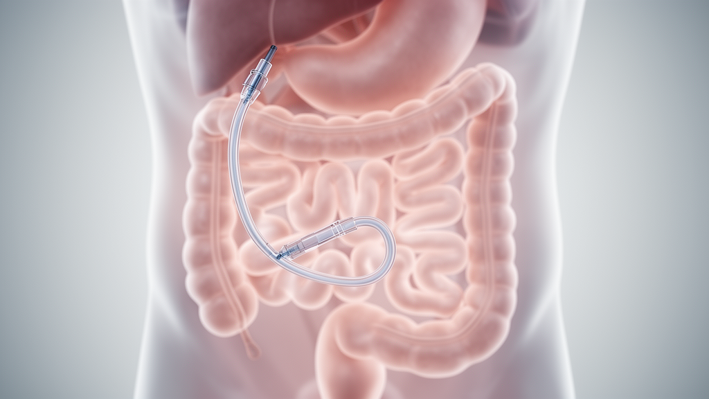
A clinical report published in Discover Medicine details the rare distal intraluminal migration of a jejunostomy Foley catheter across the ileocecal valve and into the ascending colon in a thirty-one-year-old female patient, where accurate diagnosis was established using computed tomography and successful retrieval was achieved through a distal ileal enterotomy without bowel resection. Foley catheters are occasionally employed as feeding tubes through enterostomy sites because of their broad availability, low expense, and straightforward placement, although enteral feeding through a jejunostomy is widely used to provide nutritional support following gastrointestinal procedures. While catheter-related complications typically involve tube obstruction, dislodgement, peristomal infection, and intraluminal displacement into the small bowel, distal advancement across the ileocecal valve into the large intestine represents an exceptionally rare clinical occurrence that poses distinct diagnostic and therapeutic challenges.
The patient, a female in her third decade of life without preexisting comorbidities, had previously presented with jejunal perforation and peritonitis, which had been managed surgically through an exploratory laparotomy and the creation of a double-barrel jejunostomy. On postoperative day one, a sixteen French Foley catheter was inserted through the enterostomy to establish distal enteral feeding, with its internal balloon inflated using approximately ten milliliters of sterile water. External stabilization was provided solely by a single non-absorbable suture placed at the skin exit site of the jejunostomy, without the use of an external retention disc or an auxiliary fixation device. The catheter remained in situ and functioned for approximately ten days before the patient herself noticed and reported that the feeding tube had migrated inward into the distal intestinal loop.
Despite the intraluminal displacement of the feeding tube, the patient remained clinically stable and experienced only mild abdominal discomfort without vomiting or other acute symptoms. A key clinical factor facilitating early detection without urgent decompensation was the continuous functionality of the proximal double-barrel jejunostomy stoma, which permitted uninterrupted enteral feeding and maintained the patient’s nutritional stability. Because clinical examination revealed no signs of mechanical bowel obstruction and the patient remained stable, the medical team initially adopted conservative observation over several days with the expectation that the detached catheter might pass spontaneously per rectum. However, spontaneous expulsion failed to take place, prompting progressive diagnostic evaluation to determine the precise location and trajectory of the migrated device.
Abdominal ultrasonography was performed on day ten as the initial diagnostic investigation, focusing on the evaluation of the right hypochondrium. The examination demonstrated a well-defined, rounded echogenic structure situated within the bowel lumen that corresponded directly to the inflated Foley catheter balloon, raising immediate suspicion of intraluminal migration with distal impaction. Although ultrasonography served as an effective first-line assessment to confirm the presence of the balloon inside the digestive tract, acoustic and anatomical constraints made it insufficient to delineate the complete course of the catheter along the convoluted intestinal loops, which necessitated advanced cross-sectional evaluation.
On day eleven, the patient underwent non-contrast abdominal computed tomography using a sixty-four-slice multidetector scanner, with images reconstructed at a one-millimeter slice thickness to generate multiplanar reconstructions across the axial, coronal, and sagittal planes. The resulting scans demonstrated a tubular radiopaque catheter coursing intraluminally along the distal jejunal and ileal loops before crossing directly through the ileocecal valve. The distal tip and the inflated balloon were clearly depicted lodged within the lumen of the ascending colon adjacent to the hepatic flexure, while multiplanar reformatted views mapped the continuous trajectory of the catheter from the enterostomy entrance to its colonic terminus.
Computed tomography also provided vital anatomical information regarding the condition of the surrounding viscera, confirming that the small bowel loops maintained normal caliber without upstream dilation or features of mechanical obstruction. Imaging demonstrated no bowel wall thickening, mural pneumatosis, or signs of mesenteric ischemia, and there was no evidence of pneumoperitoneum or significant free intraperitoneal fluid. Furthermore, the jejunostomy site remained fully intact without local fluid collections, peristomal leak, or adjacent inflammatory changes, confirming that the catheter had displaced entirely within the intestinal lumen without disrupting the abdominal wall or causing transmural extraluminal perforation.
From an imaging perspective, the authors noted that identifying an elongated tubular structure inside the bowel requires differential consideration of several entities, including a displaced jejunostomy or enteral feeding tube, a retained surgical drain, an ingested intraluminal foreign body, and small bowel intussusception where a tube acts as a lead point. Careful continuous tracking of the catheter along its path from the enterostomy site using multiplanar reformations permitted definitive differentiation from retained drains or intussusception. The imaging confirmed that although the ileocecal valve normally serves as a physiological barrier against retrograde reflux and luminal transit, sustained anterograde intestinal forces can facilitate transvalvular passage.
The authors analyzed several interrelated mechanical factors that likely contributed to the intraluminal migration and subsequent transvalvular displacement of the device. Primary mechanisms included anterograde peristaltic propulsion acting on the fluid-filled balloon, which functioned as an intraluminal lead point that intestinal contractions continuously pushed downstream. This propelled movement was compounded by insufficient external fixation consisting of only a single cutaneous suture without an external retention bolster, excessive initial intraluminal tube length, and possible negative intraluminal pressures or suction forces generated during active feeding cycles and normal intestinal transit.
The structural geometry of the standard Foley catheter played a decisive role in both its distal progression and its ultimate failure to exit spontaneously. While peristalsis drove the rounded balloon through the compliant ileocecal valve into the colon, the proximal end of the catheter features a wide, bifurcated configuration housing two separate openings for balloon inflation and drainage. This bulky proximal junction became impacted at the ileocecal valve, arresting further distal movement into the large bowel and precluding spontaneous expulsion per rectum, while also impeding retrograde withdrawal and limiting opportunities for endoscopic snare retrieval.
Prior to definitive surgical intervention, a gastroenterology consultation was obtained and colonoscopic-guided retrieval was attempted; however, the endoscopic procedure was unsuccessful. Consequently, operative removal became necessary, with clinicians considering either a laparoscopic approach or open laparotomy. Because the patient had undergone previous abdominal surgery for peritonitis, significant intra-abdominal adhesions were anticipated that would complicate minimally invasive manipulation, and appropriate laparoscopic resources were unavailable at the center, leading the surgical team to proceed with an open laparotomy.
On day fourteen, open surgical exploration was executed through a midline laparotomy incision, confirming the intraluminal transit of the catheter through the distal small intestine, across the ileocecal valve, and into the ascending colon where the balloon was positioned. Rather than incising the colon or manipulating the ileocecal valve directly, surgeons performed a controlled enterotomy at the distal ileum to extract the catheter, an approach chosen to minimize colonic bacterial contamination and prevent the morbidity associated with a colotomy. Following successful extraction of the intact catheter, the distal ileal enterotomy was closed in four layers using 3-0 Vicryl absorbable sutures, achieving secure closure without the necessity of any bowel resection.
The patient experienced an uneventful postoperative recovery, during which oral nutritional intake was resumed gradually and the double-barrel jejunostomy continued to function normally without complications. Monitoring identified no secondary infections, anastomotic or enterotomy leaks, or intestinal obstruction, and at a scheduled clinical follow-up conducted two weeks after the operation, the patient remained completely asymptomatic with preserved bowel function. The authors noted that the single-case nature of the report represents a study limitation that prevents broad conclusions regarding optimal universal management protocols, while underscoring that dedicated feeding tubes with tapered profiles may reduce transvalvular impaction risks compared to standard Foley devices.
Subject of Research: Distal intraluminal migration of a jejunostomy Foley catheter across the ileocecal valve into the colon diagnosed by computed tomography
Article Title: Distal migration of a jejunostomy Foley catheter across the ileocecal valve into the colon diagnosed on computed tomography: a case report
Article References: Kumar, S., Hashim, M., Tyagi, N., Puri, S., Das, D., & Goel, S. (2026). Distal migration of a jejunostomy Foley catheter across the ileocecal valve into the colon diagnosed on computed tomography: a case report. Discover Medicine, 3(1), Article 117. https://doi.org/10.1007/s44337-026-00665-x
Image Credits: AI Generated
DOI: 10.1007/s44337-026-00665-x
Keywords: jejunostomy, Foley catheter, catheter migration, ileocecal valve, computed tomography, abdominal ultrasound, distal enteral feeding, enterotomy, ascending colon, hepatic flexure, double-barrel jejunostomy, intraluminal foreign body, peristaltic propulsion, surgical retrieval, Vicryl sutures, case report
News Source: Ophelia Keating. (November 16, 2026). Computed Tomography Identifies Rare Jejunostomy Foley Catheter Migration Into Colon. Scienmag.