In the rapidly evolving fields of medical imaging and preoperative planning, a groundbreaking study has emerged that significantly enhances our understanding of segmentation algorithms used for generating CT-based meshes of liver vessels. Conducted by a team of scholars including Pedraja, Seiffert, and Corpas-del Moral, their research delves deeply into the comparative effectiveness of various segmentation methods. This research is particularly timely given the increasing reliance on three-dimensional modeling in surgical planning and intervention, which holds the potential to revolutionize liver surgeries and improve patient outcomes.
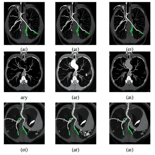
At its core, the study scrutinizes the intricacies of liver vessel segmentation by employing a range of advanced algorithms. Segmentation—the process of partitioning a digital image into multiple segments—is crucial for isolating the vessels in CT scans, enabling surgeons to visualize anatomical structures in a more intuitive manner. The authors embarked on a comprehensive evaluation of segmentation techniques to ascertain not only their accuracy but also their efficiency in clinical workflows. Such insights are vital as they inform the selection of segmentation tools, leading to more successful surgical interventions.
The study’s methodology involved a systematic approach to collecting data from multiple segmentation algorithms, applying them to a series of simulated liver CT images. By establishing a consistent testing environment, the authors could effectively compare the performance of each method under controlled conditions. This rigorous framework allowed the researchers to highlight the strengths and weaknesses inherent in each algorithm, laying a foundation for further advancements in the field of medical image analysis.
A major finding of the research is the variability in performance across different segmentation algorithms. Some algorithms excelled in delineating complex vascular structures, while others demonstrated robust performance in terms of speed and computational efficiency. This dichotomy poses critical questions for clinicians who must choose appropriate segmentation tools based on the specific demands of their practice. The study emphasizes that there is no one-size-fits-all solution; rather, the optimal choice may vary depending on the surgical context and individual patient characteristics.
To further enrich the results, the researchers employed performance metrics that included accuracy, sensitivity, and specificity. By analyzing the segmentation outputs quantitatively, the authors could provide concrete evidence of each algorithm’s efficacy. This data-driven approach not only strengthens the validity of their conclusions but also aids clinicians in making informed decisions. As surgical precision becomes increasingly paramount in complex operations such as liver resections or transplants, these insights are invaluable.
Moreover, the implications of their findings extend beyond the realm of liver surgeries alone. As technology evolves, the principles of segmentation analyzed in this study are likely to be applied to other complex anatomical regions and surgical specialties. For instance, the methods and insights can easily translate into advancements in the segmentation of brain tumors, vascular anomalies in the heart, or even orthopedic surgeries involving intricate bone structures. This versatility underlines the significance of the study within the broader landscape of medical imaging.
The research also highlights emerging trends in the integration of artificial intelligence (AI) and machine learning with traditional imaging techniques. The ability of these advanced technologies to learn and adapt from vast datasets presents new opportunities for automating segmentation processes. The authors argue that while current algorithms offer a strong foundation, the need for continuous progress in AI-powered segmentation cannot be overstated. Such technological innovations could ultimately lead to more personalized preoperative planning and reduced surgery times.
Additionally, the study draws attention to the necessity of collaboration between computer scientists, medical professionals, and engineers. In order to bridge the gap between technical algorithms and real-world surgical challenges, multidisciplinary efforts are essential. The synergy between these fields will not only drive optimization of existing techniques but will also pave the way for entirely new approaches to medical image processing. As the healthcare landscape becomes increasingly data-driven, fostering such collaborations will be vital for ongoing innovation.
Furthermore, the study invites consideration of educational initiatives aimed at empowering surgeons with knowledge of imaging technologies. As surgical techniques become more reliant on advanced imaging, it’s imperative for medical professionals to remain informed about the technology that underpins their practices. Equipping surgeons with the necessary skills to interpret these technologies effectively could lead to improved patient care and outcomes.
As a whole, this research serves as a pivotal reminder of the importance of rigorous scientific investigation in the medical field. By dissecting the capabilities and limitations of existing segmentation algorithms, Pedraja and colleagues offer critical insights that will undoubtedly inform future research directions. Their work not only enhances the current understanding of liver vessel segmentation but also sets the stage for more effective preoperative planning in various surgical disciplines.
In conclusion, the comparative study of segmentation algorithms for generating CT-based preoperative planning meshes of liver vessels marks a significant contribution to the field of medical image processing. The findings underscore the need for continuous innovation and collaboration to ensure that surgical planning can harness the full potential of advanced imaging technologies. As we move toward an era of personalized medicine, studies such as this will be instrumental in shaping the future of surgical practices, driving improvements in efficacy, safety, and overall patient outcomes.
The study stands as an exemplar of how scientific inquiry can lead to substantial advancements in clinical practice, with implications that extend well beyond the scope of liver surgeries. It calls for ongoing dialogue and cooperation among interdisciplinary teams to push the boundaries of what’s possible in medical imaging and beyond.
Subject of Research: Comparative effectiveness of segmentation algorithms for generating CT-based meshes of liver vessels.
Article Title: Comparative Study of Segmentation Algorithms for the Generation of CT-Based Preoperative Planning Meshes of the Liver Vessels.
Article References:
Pedraja, J., Seiffert, A.P., Corpas-del Moral, T. et al. Comparative Study of Segmentation Algorithms for the Generation of CT-Based Preoperative Planning Meshes of the Liver Vessels. J. Med. Biol. Eng. (2025). https://doi.org/10.1007/s40846-025-00961-4
Image Credits: AI Generated
DOI: 10.1007/s40846-025-00961-4
Keywords: CT Imaging, Segmentation Algorithms, Liver Vessels, Preoperative Planning, Medical Imaging, Machine Learning, Artificial Intelligence, Surgical Precision, Data-Driven Medicine, Multidisciplinary Collaboration.
Tags: accuracy of segmentation algorithmsadvanced imaging techniques in medicineanatomical visualization in liver surgeryclinical workflow efficiencycomparative effectiveness of segmentation methodsCT-based liver imagingdigital image partitioning in healthcareliver surgery patient outcomesliver vessel segmentation algorithmspreoperative planning advancementssurgical planning and interventionthree-dimensional modeling in surgery


