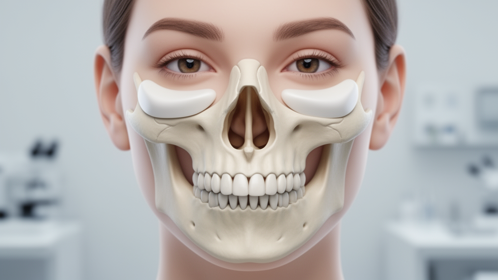
A retrospective observational investigation conducted by maxillofacial surgeons in Saudi Arabia demonstrates the clinical feasibility and preliminary safety of executing a Le Fort I osteotomy simultaneously with alloplastic midfacial implant placement during a single operative session. Published in Head and Face Medicine, the clinical study examined thirty-five consecutive adult patients presenting with combined maxillary skeletal discrepancies and upper midfacial contour deficiencies. In conventional maxillofacial workflows, corrective interventions for such deformities have historically been separated into distinct surgical stages, requiring patients to undergo standard Le Fort I maxillary advancement first, followed months later by secondary aesthetic procedures such as synthetic implant placement, structural fat grafting, or injectable soft-tissue fillers. The authors sought to determine whether consolidating skeletal repositioning and alloplastic malar and submalar augmentation into one anesthetic encounter could provide predictable structural correction without increasing perioperative morbidity, implant infection, or mechanical hardware displacement.
The anatomical rationale underlying this integrated surgical strategy addresses the intrinsic mechanical boundaries of standard maxillary orthognathic procedures. While conventional Le Fort I osteotomy reliably corrects low-level midfacial hypoplasia centered around the nasolabial region and restores balanced dental occlusion, it cannot sufficiently project deficient skeletal structures situated higher along the facial skeleton. Deficiencies involving the zygomaticomaxillary buttress, the infraorbital rim, and the lateral nasal walls remain essentially unaffected by standard horizontal maxillary advancement. Conversely, while soft-tissue augmentations like autologous fat grafting or dermal fillers can mask mild contour depressions postoperatively, the authors noted that these options remain temporary and lack the firm structural definition and long-term skeletal projection provided by solid alloplastic implants.
To evaluate the single-stage approach, the clinical investigation analyzed a cohort of thirty-five patients treated at a specialized private oral and maxillofacial hospital in Riyadh, Saudi Arabia, between December 2023 and March 2025. Conducted in accordance with the Declaration of Helsinki and reported following the Preferred Reporting of Case Series in Surgery guidelines, the protocol reviewed standardized electronic health records, operative logs, and photographic assessments from routine clinical care. The entire patient cohort was operated upon by a single experienced oral and maxillofacial surgeon assisted by a dedicated maxillofacial surgical team, an arrangement designed to ensure strict procedural consistency across every operative intervention. Because all thirty-five participants underwent bilateral midfacial augmentation alongside maxillary orthognathic correction, the investigation evaluated a total anatomical sample of seventy inserted facial implants.
The patient sample comprised thirteen male and twenty-two female individuals of Arab ethnicity, exhibiting an overall mean age of 27.77 years with a standard deviation of 4.65 years and an age range spanning from 19 to 41 years. The participants displayed diverse dentofacial discrepancies, encompassing Angle Class I, Class II, and Class III skeletal malocclusions, with several patients presenting secondary functional and aesthetic complications including vertical maxillary excess, anterior open bite deformities, and pre-existing facial asymmetry. When indicated by individual occlusal and facial planning, mandibular repositioning was performed concurrently using bilateral sagittal split osteotomy, occasionally combined with adjunctive genioplasty to achieve comprehensive lower-face harmony. Inclusion criteria required that each eligible patient undergo simultaneous maxillary osteotomy and bilateral implant insertion during a single operative session for aesthetic or functional indications, with complete perioperative and follow-up documentation available for analysis.
Preoperative digital preparation formed a fundamental component of the treatment protocol. Every surgical intervention was virtually simulated using Individual Patient Solution CaseDesigner computer software, allowing precise digital modeling of the planned maxillary bone repositioning alongside the designated malar and submalar implant footprints. Interocclusal surgical splints were fabricated prior to surgery using three-dimensional printing via sequential addition techniques, providing rigid intraoperative guidance for skeletal alignment. Preoperative radiological evaluation incorporated panoramic radiography and computed tomography scans to assess baseline maxillary osseous volume, examine anatomical contours, and exclude undetected bony or dental pathology. Patients underwent standard systemic laboratory investigations, anesthetic fitness evaluations, and routine preoperative fasting protocols, alongside intravenous prophylactic antibiotics administered immediately prior to the induction of general anesthesia.
All surgical procedures were executed under general anesthesia maintained with controlled deliberate hypotension to minimize intraoperative blood loss and optimize field visualization. The surgical team utilized a minimally invasive intraoral access approach, making a three-centimeter horizontal incision approximately three to four millimeters above the mucogingival junction extending from canine to canine. To accommodate midfacial augmentation without external incisions, the mucosal incision was intentionally extended slightly beyond standard orthognathic dimensions, facilitating subperiosteal flap elevation to expose the alveolar crest, anterior nasal spine, lateral maxillary wall, piriform aperture, and zygomatic buttress. Maxillary osteotomy cuts were executed using surgical saws and burs, and pterygomaxillary disjunction was completed utilizing a controlled twist technique. The osteotomy line was positioned slightly inferior to the traditional Le Fort I plane to maintain safe mechanical clearance between the maxillary osteosynthesis plates and the overlying cheek implants.
Through the same continuous mucosal opening, the surgical team dissected bilateral subperiosteal pockets over the malar and submalar regions according to established anatomical principles. Implant material selection was determined by preoperative digital planning, degree of skeletal atrophy, presence of facial asymmetry, and clinical judgment. Prefabricated smooth silicone templates available in small, medium, and large dimensions were utilized in thirty patients, favored for their cost-effectiveness, ease of intraoperative folding, and capacity for precise blade trimming along the infraorbital margin to eliminate soft-tissue tension. In contrast, patient-specific polyetheretherketone implants were selected for five patients who presented with severe volumetric deficits, traumatic deformities, or marked facial asymmetry. To prevent postoperative implant migration or micromotion, each implant was rigidly anchored to the underlying zygomaticomaxillary bone using two self-drilling titanium screws, consisting of an eight-to-ten-millimeter posterior screw and an anterior screw measuring approximately six millimeters placed adjacent to the paranasal region.
Prior to multilayer intraoral wound closure, the surgical fields were copiously irrigated with antimicrobial solutions containing gentamicin, metronidazole, or ceftriaxone to reduce bacterial contamination. Owing to the conservative subperiosteal dissection and meticulous intraoperative hemostasis, compressive facial dressings were omitted without precipitating hematoma formation. Postoperatively, patients completed a seven-day regimen of oral amoxicillin and clavulanic acid, coupled with regular antiseptic chlorhexidine mouth rinses. Over an average follow-up period of 14.2 months, with a median of 13 months and a total duration ranging from 6 to 24 months, all thirty-five participants achieved successful immediate skeletal repositioning and stable implant seating. Clinical photographic reviews demonstrated marked aesthetic enhancement across lateral and oblique facial profiles, and patient compliance regarding dietary restrictions, oral hygiene, and medication administration remained consistently high.
Postoperative adverse events were documented in two individuals, establishing an overall complication rate of 5.7 percent. One complication involved a localized soft-tissue infection in a female patient with a Class III skeletal pattern who had undergone simultaneous bimaxillary surgery, maxillary segmental expansion, and genioplasty; the infection developed around a silicone implant and resolved completely following targeted antibiotic therapy and surgical drainage, without necessitating implant extraction. No instances of implant exposure, persistent chronic infection, hardware loosening, or implant mobility were identified during surveillance. A binomial test comparing the observed 5.7 percent complication rate against an established historical reference benchmark of 10 percent from existing orthognathic literature yielded no statistically significant difference, demonstrating comparable clinical safety.
Statistical exploration of patient subgroups revealed a significant association between adverse events and specific surgical approaches, with adjusted residual analyses highlighting higher complication frequencies among patients undergoing segmental maxillary advancement or segmental bimaxillary procedures with genioplasty. However, the study authors emphasized that this correlation must be regarded as preliminary and hypothesis-generating rather than confirmatory due to the small total number of adverse events. Further inferential testing revealed no statistically significant associations between complication rates and implant biomaterial, participant gender, patient age, or baseline Angle skeletal classification. Despite these favorable findings, the authors outlined substantial methodological limitations, emphasizing the retrospective single-center design, the modest sample size of thirty-five patients, the involvement of a single operating surgeon, variable follow-up intervals, and the reliance on subjective satisfaction and two-dimensional photographs rather than objective three-dimensional volumetric scans. Consequently, while the single-session strategy appears feasible and eliminates secondary surgical procedures, prospective randomized trials with quantitative imaging are required to establish whether simultaneous augmentation provides demonstrable superiority over traditional staged interventions.
Subject of Research: Simultaneous Le Fort I osteotomy and midfacial alloplastic implant augmentation for midface deficiency
Article Title: Simultaneous Le Fort I osteotomy with alloplastic implant augmentation: a novel strategy for midface deficiency
Article References: Alasseri, N., AlGhamdi, K. A., AlGhamdi, M. A., & Mokhtar, E. A. (2026). Simultaneous Le Fort I osteotomy with alloplastic implant augmentation: a novel strategy for midface deficiency. Head & Face Medicine, 22(1), Article 84. https://doi.org/10.1186/s13005-026-00646-6
Image Credits: AI Generated
DOI: 10.1186/s13005-026-00646-6
Keywords: Le Fort I osteotomy, alloplastic implants, midface deficiency, maxillofacial surgery, orthognathic surgery, silicone implants, PEEK implants, malar augmentation, submalar augmentation, zygomatic deficiency, virtual surgical planning, bilateral sagittal split osteotomy, titanium screw fixation, facial aesthetics, surgical complications, case series
News Source: Ophelia Keating. (November 18, 2026). Combined Jaw Osteotomy and Cheek Implants Correct Midface Deficiency in Single Operation. Scienmag.