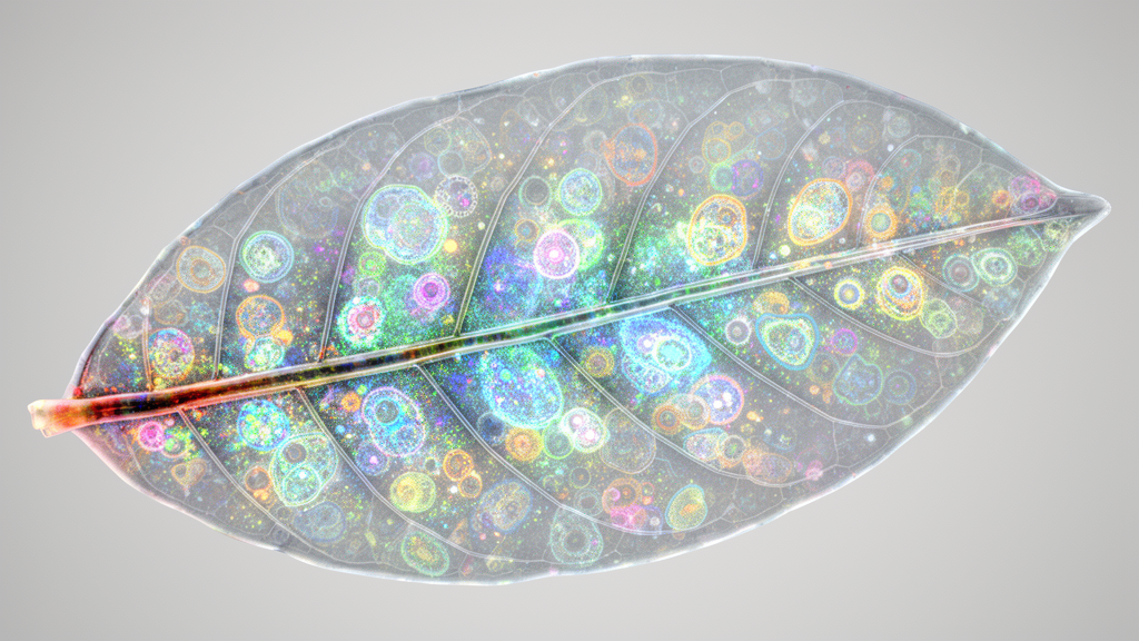
Microscopic investigation of botanical specimens has historically faced significant optical hurdles caused by the complex physical and biochemical architecture of plant tissues. Plant cells possess rigid cell walls, dense arrays of light-absorbing pigments such as chlorophyll, and high concentrations of phenolic compounds, all of which absorb and scatter light to limit optical penetration. Traditional histological approaches have resolved this obstacle through mechanical sectioning, cutting physical slices of biological structures for examination under standard slide-mounted light microscopy. Optical clearing techniques, which originated conceptually in the late nineteenth century when chloral hydrate was introduced to render plant specimens transparent, offer an alternative non-destructive path. By chemically modifying and standardizing the internal optical properties of tissues without requiring physical sectioning, modern clearing procedures enable researchers to perform three-dimensional deep optical imaging through intact, thick biological specimens.
The optical clearing process relies on a systematic sequence of chemical treatments designed to homogenize the refractive index across heterogeneous biological components. Inside an uncleared plant cell, cellular constituents exhibit widely disparate refractive indices, ranging from intra- and extracellular fluids with an index of approximately 1.35 to structural lipids at 1.47 and dense proteinaceous or structural frameworks exceeding 1.50. This pronounced refractive mismatch across cellular boundaries scatters transmitted light and severely restricts imaging depth. To overcome this limitation, optical clearing protocols typically execute four core procedural steps: chemical fixation to stabilize structural integrity, membrane permeabilization, chemical decolorization to remove light-absorbing pigments, and final refractive index matching. During index matching, endogenous aqueous fluids are evacuated or substituted with specialized clearing reagents whose refractive indices closely approximate the remaining cellular and structural matrices, thereby suppressing internal light scattering and allowing deep optical penetration into intact volumes.
Modern classification frameworks for botanical clearing protocols have evolved from early binary divisions based on organic solvent versus aqueous solution chemistry into a tripartite categorization adapted from animal tissue research. Contemporary plant clearing methods are systematically classified into hydrophobic approaches, hydrophilic techniques, and hydrogel-based systems. Hydrophobic techniques rely on non-polar organic formulations that efficiently dehydrate and clear specimens, though they often cause measurable tissue shrinkage while facilitating the visualization of larger anatomical samples. Conversely, hydrophilic protocols utilize water-soluble chemical mixtures that better safeguard native fluorescent protein signals while presenting lower chemical toxicity in laboratory workflows. Both hydrophilic and hydrogel-based strategies can induce specimen expansion, which further augments optical transparency and enhances effective spatial resolution. When integrated with advanced imaging modalities such as light-sheet fluorescence microscopy, these clearing strategies enable rapid volumetric acquisition across extensive fields of view.
Hydrophobic clearing approaches have demonstrated substantial utility in resolving complex surface structures such as foliar trichomes on medicinal and culinary herbs. In botanical analyses of oregano, Origanum vulgare, investigators deployed the specialized commercial hydrophobic clearing agent Visikol to examine protective and secretory structures across leaf surfaces. The Visikol formulation comprises a polychlorinated alcohol mixture that drives rapid tissue dehydration alongside glycerol to match the refractive index of the specimen at approximately 1.4450. When fresh oregano leaves were immersed in this solution, optical transparency was achieved within a rapid incubation timeframe of twenty to thirty minutes, presenting an efficient alternative to traditional acidified chloral hydrate in glycerol which exhibits a refractive index of 1.4280. Slide-mounted specimens cleared with this reagent allowed researchers to characterize thick-walled non-glandular trichomes aligned along the leaf veins as well as distinct capitate glandular trichomes under conventional light microscopy.
Hydrophilic clearing methodologies have proven particularly valuable for structural and developmental investigations of herbaceous model plants such as Arabidopsis thaliana. The ClearSee protocol was developed to render fixed botanical organs transparent while preserving endogenous fluorescent protein signals that are typically quenched by aggressive organic solvents. In experimental workflows using Arabidopsis seedlings, specimens were initially fixed with four percent paraformaldehyde for thirty minutes, rinsed in phosphate-buffered saline, and subsequently incubated in the ClearSee clearing solution at room temperature for seven days. The ClearSee formulation integrates ten percent weight-per-volume xylitol to assist dehydration and refractive index matching, fifteen percent sodium deoxycholate to perform membrane delipidation, and twenty-five percent urea to facilitate tissue hydration in an aqueous base. Compared to control seedlings maintained solely in saline buffer, ClearSee treatment produced comprehensive optical transparency throughout entire intact seedlings, supporting deep whole-plant fluorescence imaging.
In PEA-CLARITY protocols applied to Nicotiana tabacum leaf discs measuring seven millimeters in diameter, tissue samples were drop-fixed with an ice-cold hydrogel mixture containing four percent paraformaldehyde and four percent acrylamide, vacuum infiltrated, and thermally polymerized overnight at thirty-seven degrees Celsius. Following passive detergent clearing in sodium dodecyl sulfate for two days, the discs were incubated with an enzymatic cocktail containing cellulase and amylase for five to seven days at thirty-seven degrees Celsius. This enzymatic digestion of cellulose and starch significantly enhanced optical transparency, facilitating deep antibody penetration that permitted confocal laser scanning microscopy to detect RuBisCO within mesophyll chloroplasts while retaining nuclear-localized green fluorescent protein signals.
Rapid hydrophilic clearing has also been achieved using concentrated solutions of 2,2′-thiodiethanol, known as TDE, in the Transparent plant Organ MEthod for Imaging, or TOMEI. Developed to expedite sample preparation down to several hours, TOMEI protocols utilize 2,2′-thiodiethanol, which exhibits a high refractive index of 1.47, to clear plant tissues while maintaining fluorescent signals. The method encompasses two distinct preparation branches: TOMEI-I, which employs a fixative solution comprising acetic acid and ethanol in a one-to-three ratio, and TOMEI-II, which uses four percent paraformaldehyde in phosphate-buffered saline. When applied to two-week-old rice leaf blades approximately eighty micrometers thick, TOMEI-I fixation for one hour at twenty-five degrees Celsius followed by brief ethanol and buffer washes and immersion in ninety-seven percent TDE for twenty minutes produced optical transparency without altering cellular morphology. Subsequent confocal laser scanning microscopy clearly resolved nuclear counterstains within guard cells, mesophyll cells, and vein-associated epidermal cells in reconstructed three-dimensional volumes.
The concentration-dependent clearing dynamics of 2,2′-thiodiethanol have been systematically characterized using reproductive tissues, specifically the immature seeds of Arabidopsis thaliana. Researchers fixed immature seeds in four percent paraformaldehyde for one hour at room temperature, rinsed them in water, and systematically evaluated optical transparency across graded concentrations of aqueous TDE ranging from twenty percent to ninety-five percent alongside glycerol, water, and chloral hydrate controls. While twenty percent TDE proved functional for preserving and detecting fluorescent protein emissions, serial increases in TDE concentration progressively elevated tissue transparency until clearing efficiency reached a plateau between sixty percent and ninety-five percent. Significantly, this TDE-based protocol achieved effective optical clearing within hours, offering a rapid diagnostic workflow compared to multi-day clearing procedures such as ClearSee when rapid assessment of internal reproductive anatomy is required.
Addressing the limitations of early rapid clearing techniques, the improved TOMEI protocol, designated iTOMEI, was engineered to overcome persistent chlorophyll autofluorescence and signal attenuation of sensitive fluorescent proteins. While standard TOMEI rapidly clears structures, residual green pigments often impede fluorescence detection in highly pigmented vegetative organs. The iTOMEI procedure resolves this challenge by introducing caprylyl sulfobetaine as a specialized decolorizing detergent, buffering the solution to a weak alkaline pH of 8.0 to amplify fluorescent protein emissions, and employing a seventy-point-four percent weight-per-volume iohexol solution to provide elevated refractive index matching. In validation experiments utilizing the umbrella liverwort Marchantia polymorpha, apical thallus sections fixed in one percent paraformaldehyde were decolorized for twenty-four hours in a caprylyl sulfobetaine buffer, stained with Calcofluor White, and mounted in iohexol. Confocal laser scanning microscopy of cleared thalli successfully captured histone H2B-tdTomato fluorescent reporter signals, cleanly delineating individual fan-shaped cells and apical notch architecture.
Optical clearing techniques have likewise transformed the investigation of plant-microbe interactions by enabling three-dimensional visualization of fungal pathogens within host leaf tissues. In studies investigating bread wheat, Triticum aestivum, infected with stripe rust, Puccinia striiformis, or crown rot, Fusarium pseudograminearum, infected leaves were cleared in a solvent mixture of zero-point-fifteen percent trichloroacetic acid in ethanol and chloroform (four-to-one ratio) for forty-eight hours.
Similar clearing strategies have been adapted to track necrotrophic and foliar fungi in major crop models, including maize pathosystems colonized by Cochliobolus heterostrophus. To visualize pathogen invasion around vascular structures in Zea mays leaves, inoculated tissues were fixed in a combined glutaraldehyde, paraformaldehyde, and Triton X-100 fixative, rinsed in glycine, and stained over five days with Calcofluor White MR2 and Alexa Fluor 568-conjugated wheat germ agglutinin. The stained leaves were subsequently cleared for forty-eight hours in ScaleP, a mammalian-derived clearing formulation comprising six molar urea, thirty percent glycerol, and zero-point-one percent Triton X-100 in water.
Subject of Research: Chemical tissue clearing techniques for three-dimensional deep optical imaging in botanical research and plant-microbe interactions.
Article Title: Clearing techniques for deeper imaging of plants and plant–microbe interactions
Article References: Kim, K. W. (2024). Clearing techniques for deeper imaging of plants and plant–microbe interactions. Applied Microscopy, 54(1), Article 5. https://doi.org/10.1186/s42649-024-00098-9
Image Credits: AI Generated
DOI: 10.1186/s42649-024-00098-9
Keywords: optical clearing, plant microscopy, plant-microbe interactions, confocal laser scanning microscopy, PEA-CLARITY, ClearSee, TOMEI, iTOMEI, Visikol, ScaleP, refractive index matching, Arabidopsis thaliana, Oryza sativa, Marchantia polymorpha, Nicotiana tabacum, fungal hyphae
News Source: Denise Maddox. (November 13, 2026). Chemical Clearing Techniques Enable Deeper Three-Dimensional Imaging Across Diverse Plant Specimens. Scienmag.