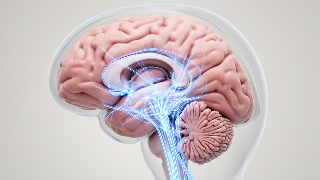
A special issue of the journal Dystonia, published in November 2026, presents a collection of studies that collectively position the cerebellum as a critical component in the distributed neural networks underlying dystonia. The editorial, authored by Linda H. Kim, Cheryl Brandenburg, and Roy V. Sillitoe from Baylor College of Medicine and Texas Children’s Hospital, synthesizes recent findings from genetic models, cellular physiology, systems-level imaging, and human electrophysiology. The central argument is that dystonia is not a disorder of a single brain region but a network phenomenon where dysfunction is distributed across the cerebellum, thalamus, basal ganglia, and cerebral cortex. This integrated view suggests that cerebellar abnormalities may contribute directly to pathological movement in some contexts, while in others, they reflect broader network dysfunction or influence both motor and non-motor features of the disorder.
The research included in this special issue spans multiple methodological approaches, offering a comprehensive look at the complexity of cerebellar involvement. Contributions range from genetic mouse models that dissect mechanistic contributions to human studies that provide critical insights into patient symptoms. This diversity of approaches allows for a cross-verification of findings, where observations in animal models guide future questions in human research, and clinical data inform the interpretation of mechanistic studies. The editors emphasize that this broad and integrated view is essential for understanding the importance and complexity of the cerebellum in dystonia, highlighting potential entry points for therapeutic development that address the disorder as a whole rather than in isolation.
Genetic mouse models have been instrumental in dissecting the mechanistic cerebellar contributions to dystonia. Adury et al. compared global Dyt1 mutations with mutations restricted to cortical and hippocampal neurons, revealing that the global mutation produces more widespread reductions in functional connectivity across sensorimotor networks. In contrast, the regionally restricted mutation generates a distinct pattern of connectivity changes in the cerebellum, striatum, and brainstem. These findings reinforce the view of dystonia as a network disorder whose expression depends heavily on the distribution of molecular dysfunction.
Complementing the work of Adury et al., Xing et al. investigated the cell-autonomous nature of Purkinje cell abnormalities. They showed that Purkinje cell-specific Dyt1 mutations do not produce significant changes in spontaneous firing and only result in subtle alterations in intrinsic physiology. This suggests that the more pronounced Purkinje cell abnormalities observed in global models may not be strictly cell-autonomous but rather dependent on circuit-level interactions. In a separate study, Xing et al. identified sex-dependent alterations in Purkinje cell firing in Sgce knockout mice, a model of DYT11 myoclonus-dystonia. Female mice exhibited more pronounced electrophysiological abnormalities, which paralleled an approximately fourfold increase in myoclonus compared with male knockout mice, although the causal relationship between these phenotypes remains to be established.
Liu et al. further examined cerebellar torsinA dysfunction using an acute, adult-onset, Cre-mediated manipulation. Reduced torsinA expression in the cerebellum decreased Purkinje cell spontaneous firing and intrinsic excitability, producing overt dystonia in the majority of animals. The emergence of severe spinning in a subset of mice raises important questions about broader network involvement, suggesting that local cerebellar dysfunction can trigger widespread motor symptoms.
Several contributions extend these mechanistic observations toward systems-level and clinical questions. Jackson et al. synthesized evidence for Purkinje cell and cerebellar nuclei dysfunction across different dystonia subtypes, highlighting impaired calcium regulation as a potential point of convergence for future investigation. Mulcahey et al. demonstrated that motor priming alters low-frequency cerebellar resting-state dynamics differently in individuals with writer’s cramp and healthy participants.
Seyyed Mousavi et al. provided rare direct electrophysiological recordings from a pediatric patient with secondary dystonia, offering a unique window into the human neural circuits involved. Task-aligned activity across basal ganglia and thalamic sites, including the ventral intermediate nucleus (VIM) and centrolateral nucleus (CL), supports the involvement of distributed cerebellothalamic and basal ganglia circuits in movement timing.
Kim et al. broadened the clinical framework by reviewing cerebellar contributions to motor and non-motor features of dystonia, particularly sleep and cognition. Iacono offered a unique perspective, linking developmental movements, adaptation to gravity, and cerebellar–basal ganglia interactions.
Collectively, the studies in this special issue position the cerebellum as an important component of the distributed networks underlying dystonia, while also emphasizing that its contribution is unlikely to be uniform across disease subtypes. Findings from genetic models show that Purkinje cell abnormalities may be direct, circuit-dependent, or modified by sex and developmental timing. Human imaging and electrophysiology further demonstrate that cerebellar function must be understood in relation to interconnected basal ganglia, thalamic, and cortical systems.
Defining when cerebellar dysfunction initiates dystonia, when it amplifies pathology elsewhere in the network, and when it can be therapeutically engaged will be essential for translating these insights into more precise and effective interventions.
The electrophysiological findings across the genetic models delineate critical distinctions between cell-autonomous dysfunction and broader network pathology. When evaluating torsinA dysfunction through an acute, adult-onset, Cre-mediated manipulation, Liu and colleagues demonstrated that reduced cerebellar expression directly decreased Purkinje cell intrinsic excitability and spontaneous firing, generating overt dystonia across most treated animals and severe spinning in a subset. In contrast, Xing and colleagues found that Purkinje cell-specific Dyt1 mutations caused only subtle alterations in intrinsic physiology without significantly changing spontaneous firing rates. This physiological divergence suggests that the severe Purkinje cell disruptions observed in global Dyt1 models do not stem solely from cell-autonomous defects, but instead require broader circuit-level dysfunction. Furthermore, the DYT11 myoclonus-dystonia model revealed that Purkinje cell firing abnormalities can be sex-dependent, with female Sgce knockout mice exhibiting more pronounced electrophysiological disruptions that coincided with an approximately fourfold rise in myoclonus compared to males, though causal links remain unproven.
Human neurophysiological and imaging investigations provide a complementary framework by identifying circuit nodes and behavioral contexts relevant to therapeutic intervention. Seyyed Mousavi and colleagues recorded direct, task-aligned electrophysiological signals across thalamic and basal ganglia targets, including the ventral intermediate nucleus and centrolateral nucleus, in a pediatric patient diagnosed with secondary dystonia. Additionally, Mulcahey and colleagues observed that motor priming modified low-frequency cerebellar resting-state dynamics differently in healthy participants compared to individuals with writer’s cramp.
Clinical and theoretical contributions within the collection further broaden the scope of cerebellar involvement beyond motor coordination. Jackson and colleagues highlighted impaired calcium regulation across Purkinje cells and deep cerebellar nuclei as a candidate molecular convergence point shared across diverse dystonia subtypes. At the systemic level, Kim and colleagues emphasized that cerebellar pathology impacts non-motor domains, specifically sleep and cognition, underscoring the clinical necessity of therapies that target overall patient quality of life. Integrating these elements with Iacono’s conceptual framework linking developmental movement, gravitational adaptation, and motor network organization, the editorial stresses that effective clinical translation will depend on resolving whether cerebellar disturbances initiate pathological signals, amplify existing network dysfunction, or serve as accessible targets for therapeutic modulation.
Subject of Research: Cerebellar involvement in dystonia networks
Article Title: Editorial: Dystonia and cerebellum
Article References: Kim, L. H., Brandenburg, C., & Sillitoe, R. V. (2026). Editorial: Dystonia and cerebellum. Dystonia, 5, Article 17463. https://doi.org/10.3389/dyst.2026.17463
Image Credits: AI Generated
Keywords: dystonia, cerebellum, neural networks, Purkinje cells, genetic models, electrophysiology, basal ganglia, thalamus, motor disorders, neuromodulation, Editorial, scientific research
News Source: Ophelia Keating. (November 9, 2026). Cerebellum Role in Dystonia Networks. Scienmag.