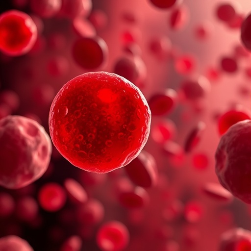
In a remarkable breakthrough that upends long-held beliefs about blood clotting, researchers at the University of Pennsylvania have uncovered a surprising player in the highly complex process of clot contraction: red blood cells. Traditionally dismissed as mere passive components trapped within clots, these cells now emerge as active agents driving clot shrinkage independently of platelets. This discovery not only challenges existing paradigms of hemostasis but also opens exciting avenues for understanding and treating clotting disorders that lead to either uncontrollable bleeding or dangerous thrombotic events like strokes.
For decades, scientific consensus held that platelets—the small, disc-shaped cell fragments circulating in the blood—were the primary force behind clot contraction. Platelets tightly pull on strands of the protein fibrin, creating a dense mesh that stabilizes the clot and stops bleeding. Red blood cells, by contrast, were thought to be bystanders, essentially filling space within the clot without influencing its mechanics. However, new experimental data from the University of Pennsylvania team reveals that red blood cells are far from inert; instead, they contribute significant contractile forces, reshaping how we understand the fundamental mechanics of blood clot dynamics.
The team’s unexpected findings came from an experiment designed to test the assumption that clot contraction requires platelets. By creating blood clots entirely devoid of platelets, researchers anticipated no contraction would occur. To their surprise, the clots shrank in volume by more than 20%. This initial observation was rigorously verified by chemically inhibiting platelet activity in normal blood samples, yet clot contraction persisted robustly. These results unequivocally demonstrated that red blood cells themselves can drive clot compaction, revealing a platelet-independent mechanism that refines the conceptual framework of thrombus formation.
.adsslot_aFbxnK0EXW{ width:728px !important; height:90px !important; }
@media (max-width:1199px) { .adsslot_aFbxnK0EXW{ width:468px !important; height:60px !important; } }
@media (max-width:767px) { .adsslot_aFbxnK0EXW{ width:320px !important; height:50px !important; } }
ADVERTISEMENT
To explain this phenomenon, the researchers enlisted the expertise of Prashant Purohit, a mechanical engineer specializing in soft materials and bioengineering. Purohit’s mathematical modeling pointed to a process known as osmotic depletion as the primary force behind the red blood cell aggregation within clots. Osmotic depletion is a physical effect typically observed in colloidal mixtures—substances where microscopic particles cluster due to imbalances in surrounding molecular concentrations. In the context of blood clots, proteins suspended in the fluid environment create an osmotic pressure gradient that effectively pushes red blood cells closer together.
As a clot develops, a fibrin meshwork forms which captures red blood cells within its intricate protein lattice. When these cells become tightly packed, proteins in the blood plasma are forced out of the narrow spaces between them, increasing the protein concentration in the surrounding fluid. This concentration difference generates a pressure that “squeezes” the red blood cells further together. Rather than being mere placeholders, the red blood cells experience a powerful external force compelling them to contract the clot volume and mechanically reinforce the fibrin network. This compelling physical interaction ensures that the clot shrinks and strengthens even in the absence of platelets pulling the fibrin strands.
Beyond osmotic depletion, the team also considered another previously proposed mechanism—bridging. Bridging involves weak molecular attractions on the surfaces of red blood cells leading to adhesion and clustering. However, Purohit’s modeling and subsequent experiments revealed that this effect is considerably weaker and insufficient to account for the observed degree of clot contraction. Experiments using modified blood samples lacking the molecules responsible for bridging still exhibited substantial clot shrinkage, but when conditions suppressed osmotic depletion forces, contraction was minimal. This elegant combination of theory and practice affirms osmotic depletion as the dominant mechanism in platelet-independent clot compaction.
This newfound role of red blood cells has critical clinical implications. Disorders such as thrombocytopenia, characterized by dangerously low platelet counts, often lead to uncontrolled bleeding due to impaired clot formation. Understanding that red blood cells can compensate, at least partially, and contribute actively to clot contraction may reshape therapeutic approaches to such conditions. Additionally, the mechanics uncovered may shed light on how clots fragment and embolize, traveling through the bloodstream to obstruct critical vessels and cause strokes. Fine-tuning the knowledge of these mechanical forces opens possibilities for novel interventions aimed at preventing or mitigating thrombotic complications.
The interdisciplinary nature of this study—melding cell biology, biophysics, mechanical engineering, and clinical insight—illustrates the power of collaboration in biomedical innovation. Using sophisticated imaging, biochemical assays, and computational modeling, the researchers unraveled complex biomechanical interactions within the clot’s microenvironment, a process that had remained elusive since blood cells were first described centuries ago. This research highlights the dynamic and mechanical contributions of each cellular player beyond their traditionally assigned roles.
From a biophysical standpoint, the study extends our comprehension of how soft, viscoelastic materials behave under mechanical stress in physiological conditions. Blood clots represent an adaptive material paradigm where cellular and protein components integrate in a delicately balanced microarchitecture. The study’s findings emphasize that cellular biomechanics—not just biochemical signaling—is instrumental in the maintenance and regulation of hemostasis. Red blood cells’ ability to aggregate and generate compressive forces challenges existing models and indicates that clot maturation is a holistic, multi-component process.
The implications also extend to biomimetic engineering and the design of synthetic materials. By understanding the natural principles governing red blood cell aggregation and fibrin network stiffening, engineers may develop advanced materials that dynamically respond to mechanical forces, with potential applications in wound healing technologies or medical devices. This insight bridges fundamental science with translational research aimed at enhancing patient care through innovative material science.
While this study marks a significant milestone, the researchers acknowledge that many questions remain. How various pathological conditions alter red blood cell mechanical properties and osmotic interactions needs further exploration. Similarly, the interplay between red blood cells, platelets, fibrin, and other blood components under diverse physiological and pathological states warrants deeper investigation. Future research will likely investigate the modulation of osmotic depletion forces as therapeutic targets and the potential for controlling clot size and stability with precision medicine approaches.
In conclusion, the University of Pennsylvania team’s discovery that red blood cell aggregation contributes actively to blood clot contraction radically changes our understanding of hemostasis. This platelet-independent mechanism, driven by osmotic depletion forces, enriches the biological narrative of how clots form, mature, and stabilize. With far-reaching implications in clinical hematology, stroke prevention, and biomaterials science, this work exemplifies the profound insights gained when engineering principles illuminate biological phenomena, ushering a new era of multidisciplinary investigation into the life-saving mysteries of blood clotting.
Subject of Research: Cells
Article Title: Red blood cell aggregation within a blood clot causes platelet-independent clot shrinkage
News Publication Date: 22-Jul-2025
Web References:
https://doi.org/10.1182/bloodadvances.2024015533
References: Blood Advances, University of Pennsylvania study supported by NIH grants R01 HL148227, P01 HL146373, R01 HL148014, R01 HL159256, and American Heart Association 25POST1357254/2025
Image Credits: Rustem Litvinov
Keywords: red blood cells, blood clots, clot contraction, platelet-independent clotting, osmotic depletion, fibrin mesh, hemostasis, thrombocytopenia, embolism, stroke, biophysics, blood biomechanics
Tags: active role of red blood cellsadvancements in medical researchblood clot contraction mechanismsblood clot dynamicschallenges to traditional blood clot theorieshemostasis and clotting disordersimplications for stroke treatmentnew discoveries in thrombosis researchred blood cells in clottingrole of platelets in hemostasisunderstanding thrombotic eventsUniversity of Pennsylvania research findings


