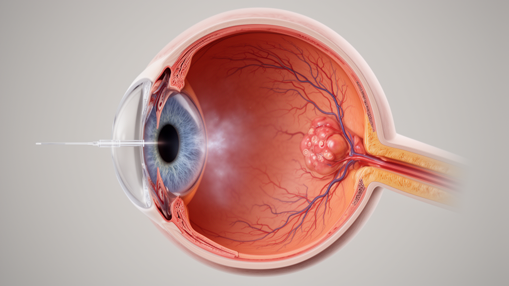
A retrospective clinical case report documents the emergence of delayed-onset hypertensive uveitis in a patient following repeated intravitreal injections of the bispecific antibody faricimab. Ten days after receiving her seventh faricimab injection, the patient presented with vision loss, mild eye pain, elevated intraocular pressure, unilateral granulomatous anterior and intermediate uveitis, and distinctive mutton-fat keratic precipitates. The clinical observation contributes new documentation to real-world safety monitoring for the medication, which has seen increasing documentation of associated intraocular inflammation in recent large clinical cohorts.
Faricimab is a humanized, bispecific monoclonal antibody designed to bind and inhibit both angiopoietin-2 and all vascular endothelial growth factor-A isoforms. Regulatory authorities have approved the therapeutic agent for treating neovascular age-related macular degeneration, diabetic macular oedema, and retinal vein occlusion.
To investigate the clinical course of the complication, investigators conducted a retrospective chart review extracting clinical data, multimodal imaging, and serial slit-lamp photography from the affected patient. The medical team applied the Standardization of Uveitis Nomenclature criteria to systematically grade the severity and location of the intraocular inflammation. Additionally, evaluators utilized the standardized Naranjo Probability Scale to formally assess the likelihood that the observed inflammatory episode represented an adverse drug reaction.
A primary novel contribution of the clinical report centers on the detailed serial photographic documentation tracking the morphological evolution and eventual resolution of the granulomatous keratic precipitates over a five-month follow-up period. The patient was successfully managed using a medical regimen consisting of topical corticosteroids alongside hypotensive eye drops, ultimately achieving complete clinical resolution of the inflammatory signs and elevated pressure.
Despite the probable attribution to faricimab therapy, the authors highlighted a significant diagnostic limitation in the retrospective review. Aqueous humor viral testing was not performed during the patient’s acute presentation, leaving a viral infectious etiology as an unexcluded alternative diagnosis that could mimic similar ocular findings.
The authors concluded that while this observation highlights a potential risk of delayed-onset intraocular inflammation, infectious etiologies must be strictly evaluated before definitively attributing such events to the therapeutic agent. The findings emphasize the vital importance of ongoing monitoring, careful diagnostic workups, and continued research into the real-world safety characteristics of faricimab in clinical ophthalmology practice.
Subject of Research: Faricimab-associated hypertensive uveitis case report and clinical evaluation
Article Title: Faricimab-associated hypertensive uveitis – a case report
Article References: Nastasi, A., Vinciguerra, R., & Romano, M. R. (2026). Faricimab-associated hypertensive uveitis – a case report. Journal of Ophthalmic Inflammation and Infection. https://doi.org/10.1186/s12348-026-00635-5
Image Credits: AI Generated
DOI: 10.1186/s12348-026-00635-5
Keywords: faricimab, hypertensive uveitis, intraocular inflammation, keratic precipitates, ophthalmology, adverse drug reaction, mutton-fat keratic precipitates, retinal vein occlusion, diabetic macular oedema, neovascular age-related macular degeneration, angiopoietin-2, VEGF-A, Naranjo Probability Scale, Standardization of Uveitis Nomenclature, corticosteroids, intraocular pressure
News Source: Ophelia Keating. (November 19, 2026). Case Report Documents Delayed Hypertensive Uveitis Following Faricimab Eye Injections. Scienmag.