A single-center retrospective study published in CVIR Endovascular reports that a calculated ratio of left adrenal aldosterone to estimated right adrenal aldosterone performs poorly for determining the subtype of primary aldosteronism. The research, led by Saachi Datta and colleagues, externally validated a method proposed by Tuandam et al. that aims to infer disease lateralization when catheterization of the right adrenal vein is unsuccessful. The study included 199 post-adrenocorticotropic hormone stimulation adrenal vein sampling cases from 2016 to 2025, with 164 patients meeting the criteria for technically successful sampling and determinate lateralization indices.
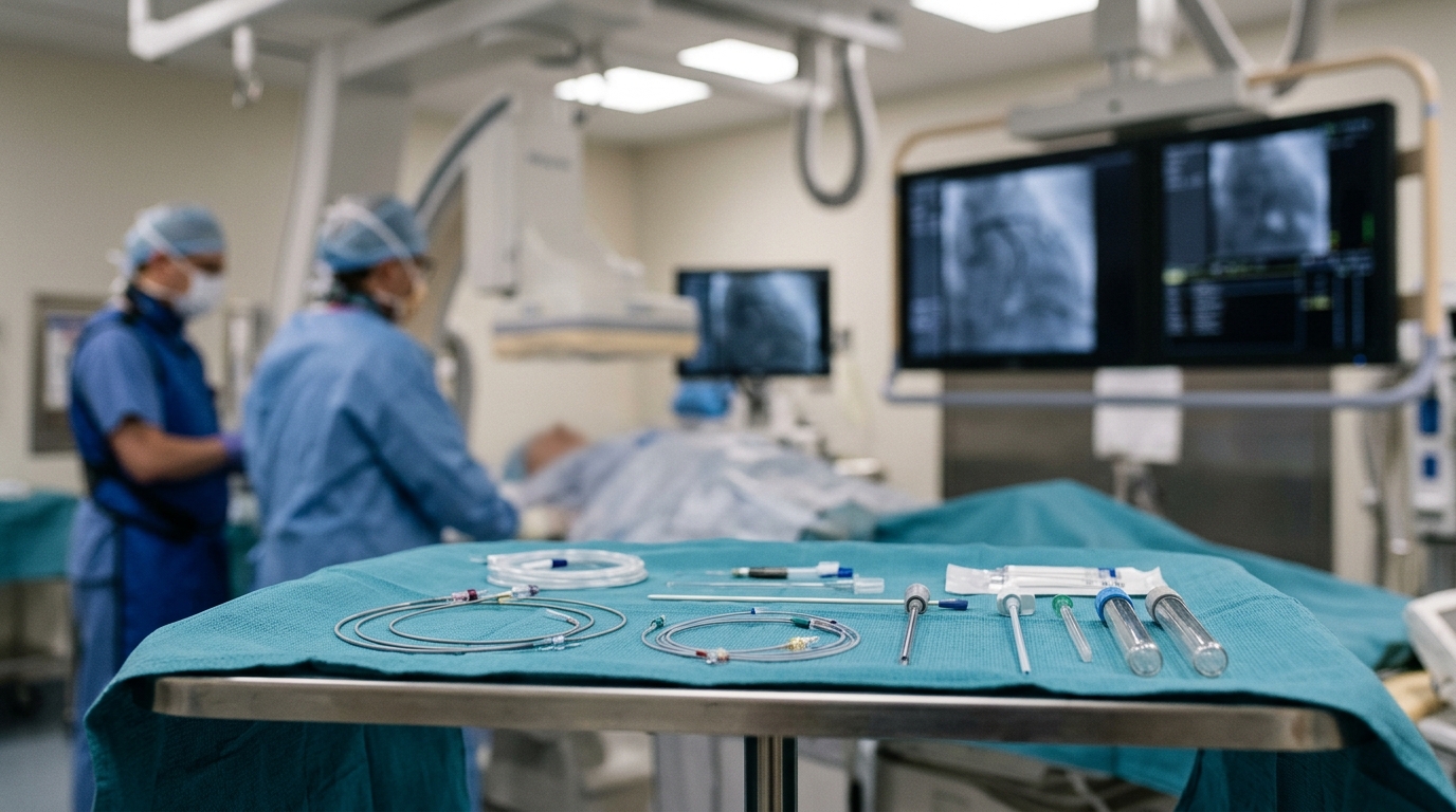
Primary aldosteronism is a common cause of secondary hypertension, occurring in up to 15% of hypertensive patients. After diagnosis, patients who are candidates for surgery often undergo adrenal vein sampling to determine whether aldosterone secretion is unilateral or bilateral. Unilateral disease typically leads to surgical intervention, whereas bilateral disease requires lifelong medical management. A common cause for technical failure during this procedure is unsuccessful catheterization of the right adrenal vein, which is challenging due to its small caliber and anatomical variation.
When right-sided sampling fails, alternative ratios have been proposed to infer lateralization using left-sided and inferior vena cava cortisol and aldosterone values. Tuandam et al. recently proposed a novel method with a published 92.6% sensitivity and 100% specificity for determining unilateral disease. This method calculates right-sided aldosterone concentration using only left adrenal vein and inferior vena cava measurements, assuming comparable cortisol production bilaterally. The ratio of left adrenal aldosterone to calculated right adrenal aldosterone is used to determine disease subtype with cutoff values of 0.33 or lower for right-sided disease, 3 or higher for left-sided disease, and intermediate values for bilateral disease.
The current study aimed to reproduce and evaluate these methods on a larger cohort at a large tertiary center. The investigators used technically successful adrenal vein sampling results as the reference standard, independent of cross-sectional imaging findings. This approach was chosen because cross-sectional imaging assesses structure rather than function, and there is a relatively high prevalence of non-functioning adrenal adenomas. By including both imaging-concordant and imaging-discordant cases, the study sought to avoid the spectrum bias that may have affected the original study, which excluded nearly one-third of its cohort for imaging-discordant results.
The final cohort consisted of 164 patients who underwent successful adrenal vein sampling for primary aldosteronism. The group included 41 patients with left-unilateral disease, 35 with right-unilateral disease, and 88 with bilateral disease. Cross-sectional imaging was concordant with the adrenal vein sampling-determined subtype in 109 of the 164 patients, or 66.5%. The mean age of the patients was 54.3 years, with 66 females and 98 males. Adrenal vein sampling was performed by five board-certified interventional radiologists using a standardized poststimulation sequential protocol.
Using the originally published thresholds of 3 for left-sided disease and 0.33 for right-sided disease, the calculated ratio correctly classified only 71 of the 164 cases. The overall accuracy for determining disease subtype was 43.3%, with a 95% confidence interval of 35.9% to 50.9%. The metric identified left-sided disease with 95.1% sensitivity and 35% specificity, right-sided disease with 71.4% sensitivity and 96.9% specificity, and bilateral disease with 8.0% sensitivity and 88.2% specificity. The area under the receiver operating characteristic curve for bilateral disease was 0.49, indicating essentially no discriminatory ability for this subtype.
To improve performance, the researchers performed receiver operating characteristic curve analysis to derive institution-optimized thresholds by maximizing the Youden’s J statistic. The optimized thresholds were 32.43 or higher for left-sided disease and 0.56 or lower for right-sided disease. Using these institution-specific thresholds, 108 of the 164 cases were correctly classified, improving the overall accuracy to 65.9%, with a 95% confidence interval of 58.3% to 72.7%. The sensitivity for bilateral disease increased substantially from 8.0% to 44.3%, while specificity for right-sided disease remained high at 95.3%.
The large shift in the optimized left-sided threshold was primarily driven by the handling of negative calculated right adrenal aldosterone values, which were replaced by an institution-derived constant of 37. This substitution directly scales the calculated ratio, making the resulting threshold inherently institution- and assay-dependent. For example, changing the constant from 37 to 100 shifted the optimal cutoff from 32.43 to 12.00 without changing overall accuracy. This finding underscores that if a calculation-based method were to be adopted, each institution would need to derive its own thresholds, requiring further validation in independent cohorts before clinical application.
Several factors may explain the differences between the original findings by Tuandam et al. and the results in this cohort. The original study had a small sample size of 68 analyzed patients, with fewer than 25% of cases having bilateral disease, which may have skewed the cutoff values. Additionally, the original study only analyzed cases with successful bilateral sampling and imaging-concordant imaging. In the current study, restricting the cohort to patients with a dominant adrenal mass concordant with the sampling-determined subtype raised accuracy with the published thresholds from 43.3% to 79.4%, approaching the originally reported performance. However, this improvement was driven by the exclusion of bilateral disease, which fell from 54% in the full cohort to 11% in this subgroup.
The equations used in the study assume symmetric cortisol secretion from both adrenal glands, yet asymmetric cortisol co-secretion may occur in up to 18% of patients. This limitation may invalidate the underlying assumption in diseased glands. A prior study by Pasternak et al. proposed an alternate index using the aldosterone-to-cortisol ratio of the left adrenal vein compared with the inferior vena cava, but this also required recalibration and institution optimization for only marginally better performance at a second center. These findings demonstrate that no calculated index has been robust enough to compare to technically successful adrenal vein sampling results.
The study has several limitations, including its retrospective design and single-center setting. Although the ratio was calculated using only left adrenal vein and inferior vena cava values, all patients in this study had technically successful bilateral sampling. Patients in whom right cannulation fails may differ in ways that affect performance, but validation in that population is precluded by the absence of a reference standard. Additionally, standard adrenal vein sampling interpretation, rather than postoperative outcome, served as the reference standard, and postoperative biochemical and clinical follow-up was incomplete for the subset of patients who underwent adrenalectomy.
The authors conclude that the calculated ratio has suboptimal performance for disease subtyping, even with institution-optimized thresholds. The index may be used as a decision-support adjunct only in limited contexts, such as implied right-sided dominant disease given its high specificity in that scenario. However, it should not replace standard adrenal vein sampling interpretation and must be institution-specific. These results reinforce the critical importance of meticulous technique when performing adrenal vein sampling to achieve technically successful bilateral cannulation and diagnostic samples, as successful cannulation remains the gold standard for determining disease subtype.
Subject of Research: External validation of calculated aldosterone ratios for subtyping primary aldosteronism
Article Title: When “right” goes wrong: poor accuracy of calculated right adrenal vein aldosterone levels in subtyping primary aldosteronism
Article References: Datta, S., Wang, R., Bhalla, V., Bahrainwala, J. Z., Seav, S., Picel, A. C., Wang, D., & Hung, M. L. (2026). When “right” goes wrong: poor accuracy of calculated right adrenal vein aldosterone levels in subtyping primary aldosteronism. CVIR Endovascular, 9(1), Article 114. https://doi.org/10.1186/s42155-026-00769-6
Image Credits: AI Generated
DOI: 10.1186/s42155-026-00769-6
Keywords: primary aldosteronism, adrenal vein sampling, aldosterone ratio, lateralization index, hypertension, endocrinology, diagnostic accuracy, adrenal gland, right, goes, wrong, poor
News Source: Ophelia Keating. (November 8, 2026). Calculated Aldosterone Ratios Show Poor Accuracy in Subtyping Primary Aldosteronism. Scienmag.