Radiographically detectable buccal bone was maintained over three years of follow-up in two adult female patients treated with an interdisciplinary orthodontic and surgical protocol, according to a retrospective case series published in BMC Oral Health. The clinical approach combined guided bone regeneration, early postoperative orthodontic activation, and the application of a modified lip bumper to address severe dentoalveolar limitations.
Managing orthodontic tooth movement beyond the alveolar envelope presents persistent challenges in individuals presenting with thin periodontal phenotypes and unfavorable muscular patterns. In such clinical circumstances, patients face an increased risk of developing alveolar defects and experiencing periodontal compromise during corrective tooth movement. The investigated interdisciplinary technique was developed as an integrated clinical protocol intended to mitigate these specific anatomical and biomechanical risks.
To evaluate the clinical application of this combined strategy over time, researchers conducted a retrospective case series involving two adult female patients who exhibited severe dentoalveolar limitations alongside a heightened risk of periodontal compromise. Dentoalveolar changes were documented longitudinally using cone beam computed tomography, with radiographic scans obtained at four distinct evaluation time points: baseline prior to intervention (T0), immediately after surgery (T1), at six months (T2), and at three years of follow-up (T3).
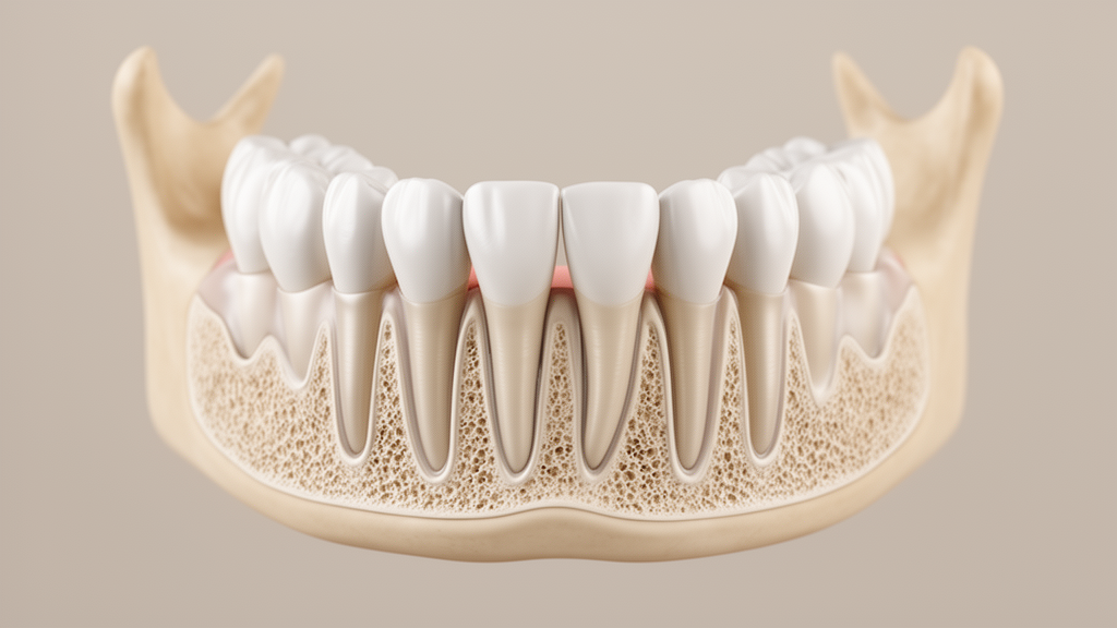
Through longitudinal cone beam computed tomography analysis, the study evaluated several anatomical parameters across the dentoalveolar complex in both patients. The investigators assessed dehiscence, fenestration, root length, buccal bone volume, and relative cone beam computed tomography grayscale values across the successive post-surgical and follow-up intervals.
Following the application of the combined clinical protocol, fenestrations in the evaluated teeth were no longer radiographically detectable within the resolution limits of the imaging protocol and did not recur throughout the three-year monitoring period. Furthermore, existing dehiscences either decreased or remained stable despite the overall magnitude of the orthodontic tooth movement performed. Buccal bone volume increased from near-zero values at baseline to measurable post-treatment levels and remained stable through the three-year follow-up examination.
The radiographic evaluations also demonstrated that relative cone beam computed tomography grayscale values declined in the immediate postoperative period before subsequently increasing at later follow-up intervals. Within the inherent limitations of a retrospective two-patient case series, the authors emphasized that the individual contribution of each treatment component cannot be isolated from these clinical observations. Furthermore, the overall effectiveness, safety, and reproducibility of the combined protocol cannot be definitively established without future prospective controlled studies using validated radiographic and periodontal outcomes.
Subject of Research: Longitudinal evaluation of guided bone regeneration and early orthodontic activation
Article Title: An interdisciplinary technique combining guided bone regeneration, early orthodontic activation, and a modified lip bumper: a two-patient case series with 3-year CBCT follow-up
Article References: García, V. J., de Blas, A. G., & Kasem, K. (2026). An interdisciplinary technique combining guided bone regeneration, early orthodontic activation, and a modified lip bumper: a two-patient case series with 3-year CBCT follow-up. BMC Oral Health. https://doi.org/10.1186/s12903-026-10459-3
Image Credits: AI Generated
DOI: 10.1186/s12903-026-10459-3
Keywords: guided bone regeneration, orthodontic tooth movement, modified lip bumper, cone beam computed tomography, alveolar envelope, periodontal phenotypes, buccal bone volume, dehiscence, fenestration, root length, grayscale values, early orthodontic activation, dentoalveolar limitations, periodontal compromise, two-patient case series, three-year follow-up, BMC Oral Health, retrospective case series
News Source: Ophelia Keating. (November 19, 2026). Buccal Bone Was Maintained Over Three Years in Two Orthodontic Patients. Scienmag.