In the realm of biological research, the meticulous study of biomolecules within intact tissue samples is an arduous endeavor that has stymied scientists for years. Researchers often grapple with the limitation of conventional imaging techniques, which can only analyze a finite number of molecules amidst the intricate landscape of cells. This challenge is particularly pronounced when it comes to studying lipid distributions, whose significance in cell function is continually being uncovered. Recent advancements in technology, particularly the innovative approach of tissue-expansion mass-spectrometry imaging (TEMI), herald a new era in this field, enabling unprecedented observation of biomolecular distributions across complex tissues such as the cerebellum.
The exploration of biomolecular localization is critical for elucidating their roles and interactions within cellular environments. Traditional imaging methods, while invaluable, suffer from restrictions that prevent them from delivering a comprehensive view of the myriad of molecules present in tissue samples. Most forms of microscopy are limited to only a handful of detectable molecules, often overlooking essential classes, particularly various lipid species. Conversely, conventional mass spectrometry excels at quantifying numerous biomolecules but typically necessitates sample destruction, thereby obscuring the spatial context required for an accurate understanding of molecular interactions.
Janelia Research Campus has witnessed a groundbreaking collaboration between two brilliant minds: Meng Wang and Paul Tillberg. Wang, a Senior Group Leader, has directed her research towards unearthing the intricacies of aging and longevity through the lens of biomolecular interactions. The challenge of visualizing these interactions within intact tissues drove her to seek innovative methodologies. Tillberg, who co-invented expansion microscopy—a technique that allows for the physical expansion of samples to unveil finer details—became a pivotal collaborator in her quest. Their combined expertise opened the door to a novel application of expansion microscopy in conjunction with mass spectrometry imaging, a fusion that holds promise for significant advancements in biomolecular research.
The novel technique developed by Wang and Tillberg involves a sophisticated method of gradually expanding tissue samples while preserving the chemical integrity of biomolecules. Unlike traditional expansion methods, which may degrade molecular constituents, their approach enables scientists to maintain the authenticity of samples, thereby providing a holistic view of molecular organization. This method allows researchers to utilize mass spectrometry imaging to identify and quantify hundreds of molecules at the single-cell level while retaining their spatial distributions within tissue samples.
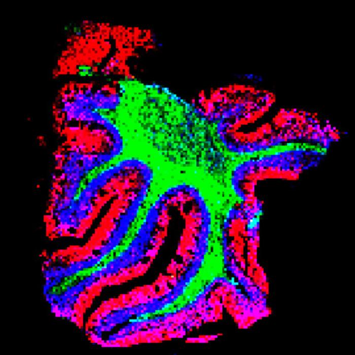
One of the most compelling applications of this new technique is its use in revealing the intricate spatial patterns of lipids, proteins, and other small molecules in the layers of the cerebellum. Previous assumptions of uniform distribution across these layers have been overturned; instead, Wang and her team have discovered distinct biomolecular signatures characteristic of each layer. This finding is revolutionary, as it indicates that the complexity of molecular distributions is far greater than previously envisioned, hinting at potential roles in cerebellar function and pathology.
Beyond the cerebellum, the applicability of this method transcends specific tissues. Wang’s team successfully adapted their approach to study biomolecular distributions in kidney, pancreas, and tumor tissues. Notably, the examination of tumor tissues revealed substantial variations in the biomolecules present, presenting vital insights into the molecular underpinnings of cancer. Understanding such variations could facilitate drug development and enhance therapeutic strategies aimed at targeting specific molecular pathways within tumors.
The potential of this technique extends far beyond its immediate applications. As scientists increasingly recognize the importance of spatial context when studying molecular biology, methods that provide high-resolution, integrated images of biomolecular distributions will be paramount. Wang and her team aim to democratize access to this technology, as it does not require specialized instruments or complex procedures, positioning it for widespread adoption across various research laboratories globally. By outlining a detailed protocol for implementing this technique, the research team hopes to empower biologists to take full advantage of mass-spec imaging in their studies.
Furthermore, this innovation could bridge a gap between the worlds of verification and visualization in biological research. With the ability to pinpoint the locations of diverse biomolecules, researchers can gain insights into the functional roles of these molecules as well as their interactions within dynamic cellular environments. The implications for our understanding of development, aging, and disease processes are profound. As biomolecular patterns can significantly influence cellular behaviors, being able to visualize these patterns can provide invaluable information regarding their functional implications.
This pioneering advancement is indicative of a trend within the field of bioscience—a transition towards multimodal approaches that leverage various imaging and analysis techniques to build a more comprehensive understanding of biological complexities. By integrating expansion microscopy with mass spectrometry imaging, scientists are not merely refining existing methods, but are crafting a new paradigm that provides clearer, more actionable data regarding the biochemical processes that orchestrate life.
The future of biomolecular research looks more promising than ever, with the potential for numerous applications arising from this groundbreaking work. As researchers continue to dissect the layers of complexity within biological systems, technologies like TEMI will become indispensable tools for unraveling the intricate web of interactions that define cellular function and behavior. Ultimately, such innovations will enhance our capacity to explore the molecular basis of health and disease, driving forward the frontiers of medical science.
Advancements in bioscience will continue to depend on the creativity and collaboration of multidisciplinary teams, much like the synergy established between Wang and Tillberg at Janelia. By fostering an environment of cooperation between disparate research fields, scientists can devise compelling solutions to some of the most pressing challenges in biological research. The integration of innovative imaging techniques not only enhances our understanding of fundamental biological processes but also accelerates the translation of this knowledge into clinical applications that may one day improve human health.
In summary, as we harness and refine these emerging technologies, we inch closer to forging a detailed and nuanced understanding of the intricate tapestry of life at the molecular level. Our ability to visualize and characterize the biomolecules that define us as living beings is crucial for unlocking the secrets of biology—secrets that, when understood, can lead to transformative breakthroughs in medicine and health.
Subject of Research: Tissue-expansion mass-spectrometry imaging (TEMI)
Article Title: TEMI: tissue-expansion mass-spectrometry imaging
News Publication Date: 22-Apr-2025
Web References: http://dx.doi.org/10.1038/s41592-025-02664-9
References: Nature Methods
Image Credits: Zhang and Ding et al.
Keywords
Research methods, Imaging, Mass spectrometry, Molecular imaging, Single cells, Tools
Tags: advancements in biological imagingbiomolecular localization techniquesbreakthrough research collaborationscerebellum biomolecule studycomprehensive biomolecule observationconventional microscopy limitationsJanelia Research Campus discoverieslipid distribution analysis in cellslipid species in cellular functionspatial context in mass spectrometrytissue expansion mass spectrometry imagingtissue sample analysis innovations


