Structural neuroimaging examinations demonstrate intracranial abnormalities in approximately sixty-two point five percent of evaluated individuals carrying genetic alterations in the Chromodomain Helicase DNA-Binding Domain 8 gene, according to an extensive systematic literature review and multi-patient clinical evaluation published in the Journal of Rare Diseases. By synthesizing neuroimaging records across dozens of published cases alongside seven newly examined individuals, the clinicians established that while central nervous system manifestations remain highly variable rather than uniform or pathognomonic, structural alterations cluster across distinct supra- and infratentorial cerebral territories. These structural findings encompass prominent Virchow-Robin perivascular spaces, asymmetric cerebellar tonsillar displacement, ventricular enlargement, and bilateral hyperintense white matter alterations that persist beyond standard chronological myelination stages.
Chromodomain Helicase DNA-Binding Domain 8 Related Neurodevelopmental Disorder with Overgrowth, designated in medical literature as CHD8-NDD, represents an exceptionally uncommon genetic syndrome documented in roughly one hundred twenty individuals worldwide. Prior clinical reports indicate that the condition arises predominantly from heterozygous de novo loss-of-function variants in the CHD8 locus situated on chromosomal band 14q11.2. The syndrome is clinically defined by primary phenotypic hallmarks including macrocephaly observed in eighty percent of patients, somatic overgrowth in eighty percent, and neurodevelopmental or neuropsychiatric manifestations such as autism spectrum disorder in seventy-five to eighty percent, developmental delay in eighty percent, and intellectual disability in seventy-five to eighty percent. Affected individuals also frequently exhibit attention-deficit/hyperactivity disorder, anxiety, sleep disturbances, gastrointestinal difficulties, muscular hypotonia, and characteristic facial dysmorphisms. Although earlier isolated reports suggested structural brain differences might occur in roughly half of diagnosed individuals, systematic synthesis addressing intracranial imaging features had remained entirely absent from the medical literature.
To rigorously delineate cranial presentations, the investigators coupled a clinical case series of six pediatric index patients and one related adult with a Preferred Reporting Items for Systematic Reviews and Meta-Analyses compliant literature review. Clinical phenotyping and whole-exome sequencing were performed for six unrelated pediatric index cases presenting to hospital clinics with developmental delays, intellectual disability, autistic traits, or overgrowth, alongside the phenotypically unaffected mother of one patient. Genetic variants in CHD8 were classified following standard American College of Medical Genetics and Genomics criteria and Association for Clinical Genomic Science protocols. Neuroimaging scans, acquired predominantly through magnetic resonance imaging across one point five Tesla and three Tesla scanners, were systematically inspected by board-certified neuroradiologists possessing over fifteen years of clinical diagnostic experience. In addition, total intracranial volumes were objectively estimated through SynthSeg software within FreeSurfer version eight point one point zero across available whole-brain sequences.
The study protocol further executed a comprehensive systematic literature screening across PubMed, Clarivate Web of Science, and Orpha.net databases covering records indexed through January 2026. The search strategy paired nomenclature for CHD8 and chromosomal region 14q11.2 with terms encompassing neuroimaging modalities including magnetic resonance imaging, computed tomography, and ultrasound. After deduplicating initial database returns and screening reference bibliographies, investigators evaluated thirty-nine candidate papers in qualitative synthesis and ultimately incorporated sixteen eligible studies providing verified genetic testing and documented cerebral imaging data. The synthesis evaluated findings from thirty-two individuals carrying isolated CHD8 variants, comparing their radiological patterns against patients harboring contiguous submicroscopic structural rearrangements involving 14q11.2. Because twelve of the sixteen incorporated studies represented uncontrolled case reports, investigators conducted descriptive analyses without formal risk-of-bias scoring tools, noting an expected high risk of bias across the retrospective literature.
Within the newly reported institutional cohort, patient one, a sixteen-year-old male carrying a heterozygous pathogenic frameshift variant designated c.3638del, p.Gly1213Aspfs*40, presented with severe developmental delays, autism spectrum disorder with verbal stereotypies, behavioral dysregulation, hypotonia, and prominent macrocephaly measuring sixty-one point six centimeters. Magnetic resonance imaging obtained at thirteen years of age identified bilateral T2-weighted hyperintense white matter signal alterations concentrated within periventricular regions, subcortical U-fibers, and deep white matter structures that could not be explained by incomplete myelination. Patient two, a seven-year-old male with a heterozygous pathogenic missense alteration designated c.5488C>T, p.Arg1830Cys, presented with nonverbal autism spectrum disorder and normocephalic cranial measurements. His brain imaging displayed matching supratentorial T2 hyperintense white matter alterations, prominent perivascular spaces, ongoing age-appropriate myelination, and a retrocerebellar infratentorial arachnoid cyst, confirming that white matter signal changes can manifest across distinct mutation classes.
The cohort also revealed critical insights regarding familial inheritance patterns and variable clinical penetrance. Patient three, a sixteen-year-old female exhibiting intellectual disability, autistic features, severe aortic arch stenosis requiring early infant surgical reconstruction, and macrocephaly, was found to harbor a heterozygous frameshift alteration designated c.3343del, p.Ala1115Leufs*39, currently classified as a class three variant of uncertain significance. Familial segregation testing identified the identical variant in her thirty-nine-year-old mother, designated patient four, who maintained entirely normal childhood cognitive development and adult psychiatric function despite having documented childhood macrocephaly. High-resolution magnetic resonance imaging revealed asymmetrical low-lying cerebellar tonsils corresponding to elevated population percentiles in both mother and daughter, alongside total intracranial volumes exceeding the ninety-seventh percentile. The authors noted that while variants of uncertain significance cannot formally define the core disease spectrum, these parallel neuroanatomical configurations support growing evidence that maternally inherited alleles may manifest with milder or subclinical phenotypes in female carriers.
Additional pediatric cases reinforced the recurrent presence of infratentorial descent and enlarged perivascular channels throughout the brain parenchyma. Patient five, a fifteen-year-old male with a class four splice variant designated c.2486+1G>A, p.?, exhibited global developmental delay, macrocephaly measuring sixty-two point eight centimeters, low-lying cerebellar tonsils at the ninety-fifth percentile, prominent perivascular spaces, a developmental venous anomaly, and an incidental Rathke cleft cyst. Patient six, an eight-year-old boy carrying a de novo class five stop-gain nonsense variant designated c.1540G>T, p.Glu514*, presented with motor coordination deficits, autism spectrum disorder, macrocephaly, prominent perivascular spaces, and tonsillar descent reaching the ninetieth to ninety-fifth percentile. Patient seven, a six-year-old girl with a de novo missense mutation designated c.3289C>G, p.His1097Asp, presented with early-onset epilepsy, developmental delays, facial dysmorphisms, and prominent perivascular spaces on neuroimaging, with automated volumetric calculations confirming an intracranial volume exceeding the ninety-ninth percentile.
Synthesizing the wider published literature, the review confirmed that objective neuroradiological abnormalities occurred in twenty out of thirty-two individuals carrying isolated CHD8 alterations, representing sixty-two point five percent of imaged patients. Prominent Virchow-Robin perivascular spaces appeared in twenty-two point six percent of individuals, while asymmetric low-lying cerebellar tonsils or Chiari type one malformations occurred in twenty-two point six percent. Ventricular enlargement, specifically ventriculomegaly, was described in twelve point five percent of cases, alongside cerebellar vermis atrophy in six point five percent and basal ganglia signal alterations in six point four percent. Although clinical macrocephaly was historically documented in roughly eighty percent of individuals, neuroimaging reports explicitly cataloged macrocephaly in only thirty-two point three percent of imaging datasets. Automated volumetric calculations performed within the authors’ clinical series indicated total intracranial volumes consistently exceeding normative pediatric percentiles, demonstrating that traditional radiologic reporting markedly underestimates actual cranial enlargement.
The investigators also contrasted isolated single-gene disruptions against broader contiguous chromosomal rearrangements spanning region 14q11.2, which encompasses both CHD8 and the adjacent chromatin regulator SUPT16H located approximately one hundred fifty kilobases proximal. Among patients harboring microdeletions of 14q11.2, neuroimaging abnormalities were identified in fifty percent of documented cases, comprising ventriculomegaly in thirty-three point three percent, cerebral atrophy in twenty percent, and posterior arch asymmetry of the dens in twenty percent. Duplications of 14q11.2 produced cerebellar vermian fissures alterations and cephaloceles, while chromosomal translocations exhibited periventricular lesions in sixty-six point six percent, pyramidal tract lesions in thirty-three point three percent, and occasional mass lesions. Patients with small marker chromosomes demonstrated cerebellar volume reductions and expanded extra-axial cerebrospinal fluid reservoirs, emphasizing that contiguous genomic rearrangements produce substantially broader syndromic manifestations than isolated single-gene haploinsufficiency.
Crucially, comparing isolated CHD8 alterations against broader 14q11.2 structural rearrangements allowed the researchers to propose specific anatomical genotype-phenotype correlations. Corpus callosum dysgenesis, frequently documented in patients with isolated SUPT16H variants, was notably absent among all reviewed individuals carrying isolated CHD8 variants. Instead, isolated CHD8 disruption appears preferentially linked to macrocephaly, lateral ventricular prominence, enlarged perivascular spaces, and downward tonsillar displacement. When chromosomal deletions or duplications capture both adjacent loci, clinical and neuroanatomical phenotypes likely represent combined gene dosage effects, complicating the attribution of specific abnormalities. Furthermore, the researchers highlighted that incidental neurovascular and sellar findings, such as developmental venous anomalies and Rathke cleft cysts, occur at rates comparable to healthy control populations and must not be misinterpreted as syndromic malformations.
The study authors highlighted several methodological limitations inherent to retrospective rare disease research that warrant cautious interpretation. The total sample size of thirty-two individuals with available neuroimaging remains modest, reflecting the extreme rarity of diagnosed cases globally. Because published literature consists predominantly of retrospective case descriptions lacking systematic control groups, publication and selection biases likely inflate reported abnormality rates above true population baseline frequencies. Furthermore, imaging acquisition protocols lacked historical standardization, relying on varied field strengths and heterogeneous sequences across different international medical centers. While automated SynthSeg volumetric modeling demonstrated acceptable concordance against three-dimensional MPRAGE scans, prospective studies utilizing standardized high-resolution sequences will be essential to establish definitive neurodevelopmental trajectories in individuals diagnosed with CHD8-related disorders.
Subject of Research: Neuroradiological findings and clinical phenotypes in CHD8-related neurodevelopmental disorder with overgrowth
Article Title: Chromodomain Helicase DNA-Binding Domain 8 (CHD8) related neurodevelopmental disorder with overgrowth: a case series and a systematic literature review on neuroradiological findings
Article References: Zerweck, L., Grundmann-Hauser, K., Kehrer, C., Grasshoff, U., Tzschach, A., Haack, T., Bevot, A., Treven, M., Pretzel, P., Hauser, T.-K., Kehrer, M., Dufke, A., Horber, V., Schuelke, M., Ernemann, U., & Bender, B. (2026). Chromodomain Helicase DNA-Binding Domain 8 (CHD8) related neurodevelopmental disorder with overgrowth: a case series and a systematic literature review on neuroradiological findings. Journal of Rare Diseases, 5(1), Article 105. https://doi.org/10.1007/s44162-026-00231-0
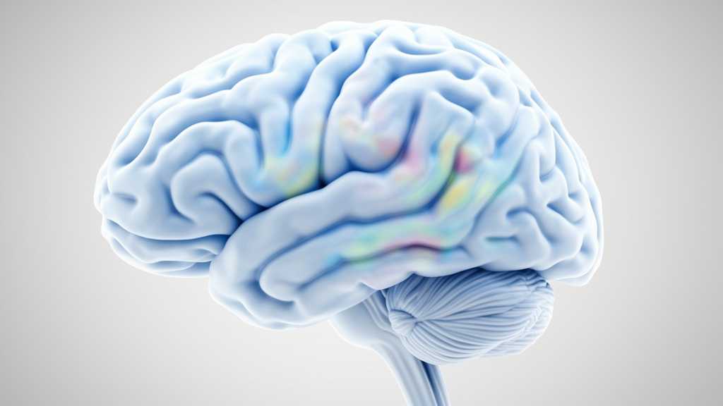
Image Credits: AI Generated
DOI: 10.1007/s44162-026-00231-0
Keywords: CHD8 gene, CHD8-NDD, neurodevelopmental disorders, overgrowth syndromes, macrocephaly, neuroradiology, magnetic resonance imaging, cerebellar tonsillar ectopia, perivascular spaces, white matter hyperintensities, ventriculomegaly, autism spectrum disorder, chromosome 14q11.2, SUPT16H gene, total intracranial volume, pediatric genetics, systematic literature review
News Source: Juliet Wilcox. (November 17, 2026). Brain Imaging Abnormalities Identified in Most Patients With CHD8 Gene Variants. Scienmag.