In recent years, pallidal neurostimulation has emerged as a transformative therapy for patients battling dystonia, a complex movement disorder characterized by sustained muscle contractions and abnormal postures. However, a newly published study unearths a paradoxical effect associated with this otherwise promising intervention. Researchers have identified bradykinesia—an insidious slowing of movement—as a side effect induced by pallidal deep brain stimulation (DBS) in certain dystonia patients. The study, led by Lange et al., offers groundbreaking insights into the clinical risk factors and the precise anatomical underpinnings of this unintended consequence, opening new avenues for personalized neuromodulatory treatments.
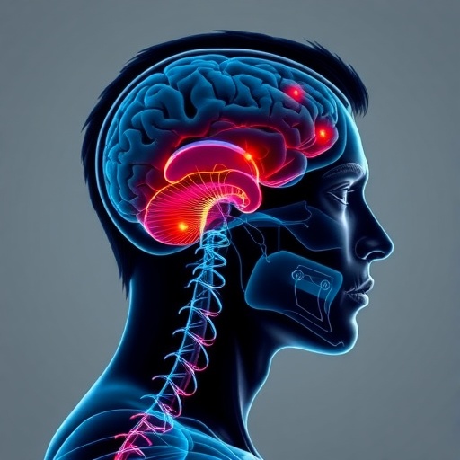
Dystonia patients often endure debilitating motor symptoms that conventional pharmacological options fail to alleviate adequately. Pallidal neurostimulation, targeting the internal segment of the globus pallidus (GPi), modulates aberrant neural circuits implicated in dystonia pathophysiology. By delivering electrical impulses via implanted electrodes, DBS aims to restore the balance of excitatory and inhibitory signaling, thus mitigating involuntary muscle contractions. Yet, despite these advancements, clinical observations have recorded instances where pallidal stimulation precipitated bradykinetic symptoms reminiscent of parkinsonism, muddying the therapeutic landscape.
The comprehensive study conducted by Lange and colleagues represents one of the first concerted efforts to elucidate the clinical determinants of bradykinesia triggered by pallidal neurostimulation. Their work involved meticulous clinical assessments combined with cutting-edge neuroanatomical mapping, illuminating how specific stimulation parameters and patient characteristics contribute to the risk profile. Notably, the researchers leveraged advanced imaging and electrophysiological data to chart the pallidal subregions most implicated in inducing motor slowing, potentially revolutionizing surgical targeting strategies.
A striking revelation from the research is the heterogeneity in patient responses to pallidal DBS, indicating that bradykinesia is not a universal side effect but rather one influenced by individual neuroanatomical and clinical factors. The authors emphasize the importance of recognizing preexisting vulnerabilities, such as subtle parkinsonian features or particular dystonic phenotypes, which may predispose patients to develop bradykinesia upon neurostimulation. This nuanced understanding challenges the prevailing one-size-fits-all approach and underscores the necessity for tailor-made therapeutic regimens.
The anatomical mapping component of the study elucidates the role of distinct GPi subterritories in modulating motor outcomes. Stimulating the posteroventral GPi, traditionally targeted for dystonia, yielded differential effects on upper versus lower limb bradykinesia, suggesting a somatotopic organization within this nucleus. Lange et al. demonstrated that inadvertent current spread to adjacent pallidal fibers might disrupt circuits essential for normal movement execution, thus provoking bradykinetic phenomena. This insight accentuates the criticality of precise electrode placement and fine-tuned stimulation parameters.
Beyond the realm of surgical planning, the study explores how stimulation settings—frequency, pulse width, and amplitude—interact with the anatomical substrate to influence side effect profiles. Modulating these parameters could mitigate the risk or severity of bradykinesia, presenting clinicians with a repertoire of adjustable levers to optimize patient outcomes. The dynamic relationship between stimulation intensity and bradykinesia is reminiscent of a delicate neurophysiological balance, where tipping points may inadvertently suppress normal motor function.
Clinically, the findings have profound implications for patient counseling and postoperative management. Awareness of bradykinesia as a plausible complication enables healthcare providers to detect early motor slowing symptoms and adjust DBS settings proactively. Moreover, identifying patients with risk factors—such as older age, longer disease duration, or concomitant parkinsonian signs—can inform preoperative discussions on expected benefits and potential trade-offs, fostering shared decision-making grounded in transparency.
This research also sheds light on the broader neurobiological interplay between dystonia and parkinsonism, two disorders traditionally viewed as distinct yet increasingly recognized to share overlapping pathophysiological elements. The induction of parkinsonian bradykinesia by pallidal stimulation for dystonia challenges binary diagnostic frameworks and prompts reconsideration of basal ganglia circuit dysfunctions. It evokes questions about the adaptability of motor networks under electrical modulation and the fine line between therapeutic and adverse effects.
Importantly, the study’s methodology reflects an integrative, multidisciplinary approach, combining clinical neurology, neurosurgery, neuroimaging, and computational modeling. Such holistic strategies are pivotal in unraveling the complex brain network dynamics involved in movement disorders and their modulation. The incorporation of patient-specific imaging data empowers surgeons to tailor electrode trajectories with unprecedented precision, minimizing off-target effects and enhancing efficacy.
Looking forward, the study by Lange et al. paves the way for future research endeavors aiming to refine neuromodulation technologies. Innovations such as directional leads, closed-loop stimulation, and adaptive DBS systems, which respond in real-time to neural activity patterns, could reduce adverse effects like bradykinesia. Furthermore, expanding the clinical dataset with longitudinal monitoring may clarify the trajectory and reversibility of stimulation-induced motor slowing, enriching the therapeutic index of pallidal DBS.
In conclusion, the discovery of bradykinesia induced by pallidal neurostimulation in dystonia patients encapsulates the complexities inherent in neuromodulatory therapies. It underscores that despite remarkable advances, the brain’s intricate circuitry demands respect and precise intervention. The implications extend beyond dystonia, offering insights relevant to a spectrum of neuropsychiatric conditions treated with deep brain stimulation. As the field evolves, balancing symptom control with preservation of natural motor function remains the paramount objective.
This study is a testament to the ongoing quest for optimized, individualized treatment paradigms in neurology. It calls for clinicians and researchers alike to remain vigilant about emerging side effects and to harness technological innovations to enhance safety and efficacy. By deepening our understanding of how pallidal neurostimulation intersects with basal ganglia networks, we edge closer to unlocking tailored therapies that can transform the lives of patients burdened by disabling movement disorders.
The findings also highlight the essential role of patient-centered care, where detailed phenotyping and risk stratification inform nuanced treatment choices. As neurostimulation technology becomes increasingly sophisticated, integrating clinical, neuroimaging, and electrophysiological data will be crucial to minimize adverse events and maximize therapeutic gain. Ultimately, this research reaffirms the delicate balance between alleviating pathological motor symptoms and preserving the integrity of normal motor pathways.
As the neuroscience community digests these insights, the ripple effects are likely to spur innovation in device design, surgical technique, and postoperative management protocols. The paradigm is shifting from uniform stimulation approaches to personalized neuromodulation strategies, heralding a new era in functional neurosurgery. Studies such as this underscore how iterative feedback between clinical practice and research propels progress and catalyzes breakthroughs.
In sum, bradykinesia induced by pallidal stimulation delineates a critical frontier in movement disorder therapy. Through meticulous clinical investigation and sophisticated anatomical mapping, Lange and collaborators have charted a path toward safer, more effective neuromodulation for dystonia. Their work embodies the spirit of translational neuroscience—bridging bench to bedside—to unlock novel, life-changing treatments for patients contending with complex, refractory neurological diseases.
Subject of Research: Bradykinesia induced by pallidal neurostimulation in dystonia, clinical risk factors, and anatomical mapping
Article Title: Bradykinesia induced by pallidal neurostimulation in dystonia: clinical risk factors and anatomical mapping
Article References:
Lange, F., Guarin, D.L., Mosert, S. et al. Bradykinesia induced by pallidal neurostimulation in dystonia: clinical risk factors and anatomical mapping.
npj Parkinsons Dis. 11, 308 (2025). https://doi.org/10.1038/s41531-025-01177-8
Image Credits: AI Generated
Tags: bradykinesia as a side effectclinical determinants of bradykinesiadeep brain stimulation risksdystonia treatment advancementselectrical impulse therapy for dystoniaglobus pallidus internal segmentmovement disorder therapiesneuromodulatory treatment optionspallidal neurostimulationparkinsonism-like symptoms in dystonia patientspersonalized treatment for dystoniaunintended consequences of DBS


