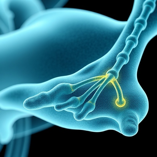
In a groundbreaking stride for regenerative medicine, researchers at Xi’an Jiaotong University have unveiled a revolutionary technique that harnesses electrohydrodynamic (EHD) bioprinting to produce living skeletal muscle tissues with unprecedented cellular alignment. This innovation promises to bridge a critical gap that has long stymied tissue engineering: replicating the intricate internal structure of real muscle, where myofibers are meticulously ordered to ensure optimal strength and function.
Traditional efforts to fabricate functional human muscle in the lab have grappled with the complexity of muscle architecture. While it is possible to shape tissues externally into muscle-like forms, the internal cellular organization rarely mirrors the natural orientation essential for muscle contraction and efficiency. This disparity hinders the performance of engineered muscles, leaving them structurally compromised and less functional than their biological counterparts.
The team’s novel approach leverages the physics of electrohydrodynamics—a process where a strong electric field is employed to draw out ultra-fine liquid jets, vastly enhancing the resolution of bioprinting beyond what conventional nozzle extrusion methods can achieve. Yet, high-definition printing alone was insufficient; the challenge lay in coaxing encapsulated cells to orient themselves within the printed matrix in a manner faithful to native muscle tissue.
The pivotal breakthrough came with the reimagining of the bioink formulation. By integrating alginate—a biocompatible, gel-forming polymer frequently used in bioprinting—with fibrin, a naturally occurring protein integral to blood clotting and tissue repair, the researchers exploited fibrin’s unique electrical responsiveness. During the printing process, intense electric forces elongate and align fibrin molecules within the hydrogel, reshaping them from random clusters into uniform nanofibers that trace along the direction of the printed filament.
This reorganization is precisely timed at the Taylor cone stage of printing, occurring under a high-voltage environment near 3,000 volts. Here, the synergy of electrical and mechanical forces restructures fibrin into nanoscale fibers aligned uniformly, creating a microscopic scaffold that cells instinctively follow. This means that instead of merely residing within the matrix, muscle cells are guided to orient and fuse along these nanofibers, mimicking the physiological architecture essential for functional muscle.
Dr. Ayiguli Kasimu, the study’s lead author, describes this process as “building a nanoscale road system” where the electric field is an invisible architect guiding cellular growth along desired trajectories. Because the alignment emerges intrinsically during bioprinting, the technique affords remarkable versatility. By modulating the printer nozzle’s path, the team achieved diverse fiber configurations—from linear bundles to curved and circular formations—closely replicating the myriad fiber orientations found across different human muscles.
Seeking to enhance the functional fidelity of these constructs, the researchers further enriched the bioink with conductive polymers. Skeletal muscle relies heavily on electrical signaling for synchronized contraction, and these conductive additives endowed the printed tissues with the capacity to transmit bioelectrical impulses effectively. This functional augmentation supported not only superior electrical properties but also more robust muscle cell development. Muscle fibers matured more efficiently, exhibiting heightened expression of proteins specific to muscle functionality.
The ultimate test of this technology was its performance in living organisms. Implanted into animal models bearing muscle defects, the bioprinted, aligned, and electrically conductive muscle tissues demonstrated remarkable survival, integration, and support for new muscle growth. Critically, these constructs translated into significant improvements in muscle function, signaling a major advance toward clinical applications for muscle repair and regeneration.
Beyond the immediate realm of muscle tissue engineering, this study redefines the role of electric fields in tissue fabrication. It reveals a powerful paradigm where electrical stimuli act as design signals, orchestrating the biochemical and biomechanical milieu to dictate cellular organization organically. The alignment effect stems from a dual mechanism: electrically induced migration of fibrin molecules and the mechanical stretching of the bioink during printing, both of which converge to sculpt an organized, cell-friendly environment.
Despite these promising results, the team acknowledges that many questions deserve further exploration. The detailed molecular pathways by which fibrin responds to electric stimulation remain to be fully elucidated. Moreover, optimizing parameters such as cell density, biomaterial chemistry, and long-term construct stability will be essential to translate this technology from the lab bench to therapeutic reality. Nevertheless, the conceptual leap represented by this work is clear and compelling.
By transforming the electric field from a mere printing force into a biological architect, Xi’an Jiaotong University investigators have charted a path that could revolutionize how living tissues are constructed. If successfully adapted to other organ systems, this electrohydrodynamic alignment strategy offers a scalable solution to the longstanding challenge of marrying shape with biological function in bioprinting, propelling regenerative medicine closer to the goal of fully functional organ and tissue replacements.
This research not only opens new vistas for muscle repair but also ignites a broader conversation about the intimate interplay between physical forces and biological patterning. It suggests a future where electrical cues might be routinely employed to engineer complex tissue architectures in vitro, offering unprecedented control over the form and function of lab-grown organs. The implications reach far beyond muscle, hinting at transformative possibilities across the fields of biofabrication, developmental biology, and therapeutic design.
As the field progresses, the integration of electrohydrodynamic bioprinting with advanced biomaterials and cell biology holds promise for creating living tissues that do not merely resemble their natural counterparts but function indistinguishably. This work stands as a testament to the power of interdisciplinary innovation, marrying engineering principles with cellular sciences to solve one of the most intricate puzzles in tissue engineering.
Amidst growing global demand for tissue replacements and regenerative therapies, this electrohydrodynamic bioprinting method represents a beacon of hope and a tangible step forward. By guiding cells through a carefully constructed electromagnetic landscape, researchers have harnessed a fundamental physical force to instruct biology itself—a strategy that may ultimately redefine how we build living matter on demand.
Subject of Research: Electrohydrodynamic bioprinting to align cell-laden fibrin-alginate hydrogels for skeletal muscle tissue engineering.
Article Title: Electrohydrodynamic bioprinting-induced orientation of cell-laden fibrin-alginate hydrogel for highly-aligned skeletal muscle constructs.
News Publication Date: 13-Mar-2026.
Web References:
International Journal of Extreme Manufacturing
Article DOI: 10.1088/2631-7990/ae3923
Image Credits: Ayiguli Kasimu, Zijie Meng, Zhennan Qiu, Yabo Zhang, Lang Bai, Xiao Tan, Ziyu Wang, Rosen Zhao, Qianxi Gao, Hui Zhu, Zhanguo Tong, Wurikaixi Aiyiti, Dichen Li, and Jiankang He.
Keywords: Electrohydrodynamic bioprinting, skeletal muscle engineering, fibrin-alginate hydrogel, cellular alignment, tissue regeneration, conductive polymers, bioelectrical signaling, regenerative medicine, nanofiber orientation, muscle tissue fabrication.
Tags: advanced cell encapsulation methodsbioink development for musclebiomimetic muscle structurescell alignment in bioprintingelectrohydrodynamic bioprintingfunctional muscle tissue fabricationhigh-resolution bioprinting techniquesmuscle tissue engineeringmyofiber orientation replicationregenerative medicine innovationsskeletal muscle regenerationtissue engineering challenges

