
Credit: David Belnap, Jorg Voteler
The University of Utah is one of just five institutions in the world to be awarded a $2.5 million grant to purchase a state of the art cryo-electron microscope (cryo-EM), the Beckman Foundation announced today. The microscope, which will be able to visualize the structure of proteins and DNA at an atom-by-atom scale, will be installed in the Crocker Science Center, currently under construction on Presidents Circle. The microscope's resolution is fine enough to see details such as the double-helix and ladder structure of DNA, said biochemistry professor Wesley Sundquist.
"Biochemistry and molecular biology take place on the atomic scale, enzymes work on the atomic scale, drugs bind on the atomic scale – so that's information that's really critical for understanding how biological processes work," Sundquist said.
Within the cells of our body are dynamic worlds that have remained largely enigmatic because they are so small. Though much of the cell's machinery is only a few billionths of a meter in size, they are essential. For instance, proteins work together to literally build the fibers of our being, to defend us from foreign invaders that cause disease, and to shape our DNA blueprints throughout life.
But as they say, a picture is worth a thousand words. With the specialized microscope, scientists can see the miniscule machines with their own eyes, directly documenting what they look like and how they work.
Before cryo-EM microscopy, researchers hoping to discern the atomic structure of proteins, molecules, drugs or DNA relied on X-ray crystallography, a labor-intensive process that required growing crystals of pure samples of the protein or molecule.
In cryo-EM, samples are not stained, treated or enclosed, as they are in traditional microscopy or in X-ray crystallography. Instead, they are frozen instantly. Further, the sample does not need to be as pure as in X-ray crystallography. The microscope takes many two-dimensional images of a molecule or protein that are then computationally combined to create a three-dimensional image. The new instrument will be more powerful and precise than the U's current cryo-EM unit, which is housed in the Aline Wilmot Skaggs Biology Building.
The new instrument is an FEI Titan Krios, and is scheduled to begin installation in November 2017, shortly after completion of the Crocker Science Center. Because the instrument must be assembled on site, installation will take two to three months.
The microscope's extreme sensitivity dictated the design of the room it will eventually occupy. "We need the instrument to have stable electronics and operating temperature and to hold the specimen very still. The instrument's components do these things very well," said David Belnap, director of the Electron Microscopy Core Laboratory.
The temperature in the room will be held to daily fluctuations of no more than 0.8 degrees Celsius (1.4 degrees Fahrenheit), Belnap says. The ventilation system is designed to avoid blowing on the microscope, since air breezes could disturb it. Even talking in the room during microscope operation would produce undesirable air movement and vibration, so operators will control the microscope from a separate room.
Further, to isolate the instrument room from all other vibrations in the building, the microscope will sit on a specially-designed concrete subfloor pad.
"Proteins form complexes in cells that perform the functions that are necessary for life," Belnap said. "If we need energy, proteins catalyze chemical reactions. If we need DNA copied, proteins catalyze that. The new instrument will enable us to study these structures at a level of detail that has not previously been possible."
Researchers from across campus came together to complete the grant application. Below are three examples from U researchers of the type of research that a cryo-EM microscope makes possible.
Unmasking a "Silent Killer"
Bedecked with twirls and curlicues, the festive image of a protein called PKD2 downplays the serious reason for creating it. The cryo-EM-made image reveals how specific mistakes in the precisely tousled protein triggers polycystic kidney disease, the most common inherited kidney disorder. Dubbed the "silent killer", patients with the disease often don't know they have it until they are well into adulthood. And once they know, there's not much that can be done. Based on the near atomic-resolution depiction, University of Utah scientists determined why the majority of disease-causing mutations are so dire. Published in the journal Cell, they surmised that the mistakes would cause the protein to fall apart, and cease to function. Aided greatly by the new information, they are now searching for pharmacological agents that block PKD2. They're also continuing to leverage the power of Cryo-EM to unmask a second cause of polycystic kidney disease, PKD1.
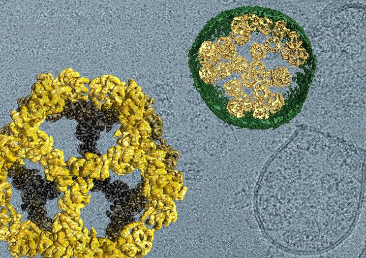
Cell Service: Micro-Delivery Systems for Targeted Treatments
Like a micro lunar lander, soccer ball-shaped "nanocages" were designed to emerge from cells, traveled through space, docked onto another bank of cells, and empty their contents. The miniature journey was documented by cryo-EM imaging and published in the top-journal Nature last year. Scientists from the University of Utah and University of Washington had developed DNA blueprints that instruct human cells to assemble the micro delivery system, inspired by the way viruses spread infection. Though the distance traveled was microscopic, the implications could be significant. U investigators are using Cryo-EM to evaluate potential applications, including for delivering drugs and therapeutics to specific sites within the body.
Caught in the Act
The scientists could hardly believe their eyes when they witnessed a molecular feat that defied their imagination, an accomplishment made possible by cryo-EM. Textbook science dictates that DNA spells out the instructions for making proteins, tiny machines that do the bulk of the work in our body's cells. What the multidisciplinary team from the University of Utah, University of Washington, and University of California, San Francisco saw instead were proteins making proteins, independent of a DNA blueprint. Think of an auto assembly line that makes cars without using instructions. The haphazardly made automobile simply doesn't work. But in the case of the instruction-free protein, the odd product might have a specific purpose , the scientists published in the journal Science. It could be part of a quality control mechanism that signals when the assembly line is not working properly. The scientists are now investigating whether defective quality control could be at least partly responsible for neurodegenerative diseases such as Alzheimer's, Amyotrophic lateral sclerosis (ALS), and Huntington's.
###
Media Contact
Julie Kiefer
[email protected]
801-597-4258
@UofUHealth
http://healthsciences.utah.edu/
############
Story Source: Materials provided by Scienmag


