A comprehensive systematic review evaluating language preservation in patients undergoing awake brain surgery has found that early postoperative language impairments are frequent but temporary, with most deficits resolving within three months of tumor resection. Published in the Chinese Neurosurgical Journal, the investigation synthesized clinical data from ten studies encompassing 532 adult patients who underwent awake craniotomy with direct electrical stimulation for diffuse gliomas in language-eloquent cerebral regions. The researchers observed that while the immediate incidence of postoperative language impairment varied widely depending on how rapidly assessments were conducted following surgery, persistent language deficits documented at three months or longer remained low across unselected cohorts, ranging between zero and 5.6 percent when evaluated using standardized whole-cohort assessment instruments. These results reinforce awake craniotomy with direct electrical stimulation as the primary surgical standard for balancing maximal tumor cytoreduction against the preservation of linguistic capabilities, while demonstrating that acute postoperative declines reflect transient network disruptions rather than irreversible damage.
Diffuse gliomas represent primary central nervous system malignancies that originate from neuroglial stem cells and exhibit a pervasive, infiltrative growth pattern throughout adjacent brain tissue. In adults, these neoplasms constitute the most frequent malignant primary brain tumors, accounting for approximately eighty percent of the twelve thousand new malignant primary brain tumors diagnosed each year in the United Kingdom. Clinical decision-making is heavily influenced by the 2021 World Health Organization classification system, which incorporates molecular markers such as isocitrate dehydrogenase mutation status along with codeletion of chromosomes 1p and 19q. While greater extent of resection is consistently associated with improved progression-free survival, overall survival, and postoperative quality of life, complete tumor removal presents acute hazards when gliomas infiltrate eloquent perisylvian pathways. Neurosurgeons must continuously balance aggressive cytoreduction against functional preservation, because severe permanent language impairments such as aphasia inflict devastating detriments on personal independence, occupational functioning, and social reintegration.
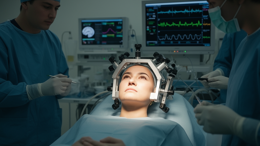
To mitigate the risk of permanent neurological morbidity, awake craniotomy paired with direct electrical stimulation mapping has emerged as the definitive intraoperative methodology for identifying functional boundaries in real time. During these neurosurgical procedures, surgeons apply localized high-frequency or low-frequency electrical currents to exposed cortical surfaces and underlying subcortical white matter tracts while the conscious patient performs standardized linguistic tasks. If the electrical current transiently disrupts verbal performance, generating speech arrest, paraphasic errors, or anomia, the stimulated site is marked as functionally critical and surgically spared. Non-invasive preoperative modalities, including functional magnetic resonance imaging, magnetoencephalography, diffusion tensor imaging tractography, and navigated transcranial magnetic stimulation, provide helpful anatomical guidance but lack the temporal precision and physiological specificity delivered by direct intraoperative stimulation mapping. Consequently, direct electrical stimulation remains the gold-standard mapping technique, enabling surgical teams to establish individualized functional margins at both cortical boundaries and deeper subcortical crossroads where infiltrative tumor borders directly abut eloquent neural circuitry.
The systematic review was prospectively registered on the PROSPERO international database under identifier CRD420261342690 and executed in accordance with Preferred Reporting Items for Systematic Reviews and Meta-Analyses guidelines. The investigators searched four major biomedical databases, comprising PubMed, MEDLINE via Ovid, Embase via Ovid, and the Cochrane Central Register of Controlled Trials, capturing literature published from 2010 through March 2026. Eligible studies encompassed randomized controlled trials, prospective cohort studies, retrospective cohorts, and clinical case series investigating adults aged eighteen years or older with supratentorial diffuse gliomas in the language-dominant hemisphere. Independent paired reviewers conducted study selection and data extraction, utilizing Joanna Briggs Institute critical appraisal checklists to evaluate methodological quality and risk of bias across individual domains. Due to extensive clinical and methodological heterogeneity regarding study designs, surgical protocols, cognitive testing batteries, and follow-up schedules, the authors precluded quantitative meta-analytic pooling, presenting instead a narrative synthesis structured around prospectively defined early, intermediate, and late follow-up observation intervals.
The ten eligible studies identified across the global literature were published between 2011 and 2025, enrolling cohorts ranging in size from seven to 225 patients, with male participants comprising 55.3 percent of the combined population. Primary tumors were predominantly located within the frontal lobe, though several cohorts also described temporal, parietal, and insular involvement. Methodological appraisal categorized seven studies as case series and three as cohort investigations, with the majority exhibiting moderate overall risk of bias due to inconsistent baseline reporting and follow-up attrition. Although the review intended to emphasize lower-grade gliomas, the analyzed population spanned the full adult diffuse glioma spectrum, encompassing World Health Organization grades 2 through 4. Higher-grade lesions predominated in multiple cohorts, with at least 114 patients across the included studies harboring confirmed grade 4 disease, including substantial representation in cohorts reported by Li, Kahana, and Vassal. Multi-grade reporting across historical classification systems ensured that high-grade infiltrative tumors contributed meaningfully to the synthesized clinical outcomes.
Early postoperative language assessments revealed marked variability across the literature, with reported deficit rates spanning an expansive spectrum from 2.2 percent to 100 percent. The researchers discovered that this apparent disparity was intimately linked to the precise timing of clinical evaluation relative to surgical completion. Investigations that assessed patients immediately following craniotomy documented exceptionally high rates of temporary linguistic dysfunction, including 80 percent in Vassal’s series and 100 percent in a cohort of seven patients evaluated by Saito and colleagues. Conversely, studies that timed early postoperative assessments between day seven and the completion of the first postoperative week reported substantially lower deficit frequencies, such as 22 percent in Saito’s 2016 cohort and 14.6 percent in Li’s cohort. Across all cohorts, the observed acute disturbances encompassed naming failures, motor aphasia, verbal repetition errors, and apraxia of speech, reflecting widespread transient physiological disruptions driven by acute perioperative cerebral edema, surgical manipulation, and temporary local vascular or metabolic perturbations.
Evaluation of late postoperative outcomes revealed a consistent clinical trajectory characterized by marked functional recovery over subsequent weeks and months. Among the four investigations that formally tracked patients at or beyond the three-month milestone, persistent language impairment remained remarkably uncommon in whole-cohort assessments utilizing standardized testing tools. In a large cohort of 225 patients evaluated by Li and colleagues, zero persistent deficits were observed at ninety days, while Sellier and colleagues similarly reported zero persistent language deficits among 81 patients followed out to one year postoperatively. In the cohort reported by Saito in 2016, only a single individual among eighteen participants exhibited persistent dysfunction at late follow-up, yielding a modest upper-bound persistent deficit rate of 5.6 percent. Intermediate follow-up studies further corroborated this rapid resolution pattern, with Vassal documenting complete functional normalization within one month, and Tsuzuki recording that patient working memory and verbal fluency fully normalized within three months following resection.
In sharp contrast to the favorable recovery observed across broader cohorts, specific subcortical tract dissections were linked to distinct, prolonged neuropsychological deficits, establishing subcortical mapping as an indispensable surgical safeguard. In a landmark cohort of 44 patients evaluated by Papagno and colleagues, patients who underwent surgical resection involving the uncinate fasciculus exhibited an alarming 47 percent rate of persistent impairment on a standardized famous face naming task at three months post-surgery. By comparison, individuals whose uncinate fasciculus was anatomically preserved exhibited an impairment rate of only 10 percent on the identical naming battery. The authors emphasized that this 47 percent figure represented a highly specialized proper-naming deficit within an anatomically targeted subgroup rather than generalized communicative failure. Nevertheless, the finding underscores that functional preservation cannot be secured by cortical mapping alone, highlighting the critical imperative to incorporate connectomic principles that actively track and safeguard deep associative white matter tracts, including the uncinate fasciculus, arcuate fasciculus, and inferior fronto-occipital fasciculus.
The synthesis also highlighted substantial confounding factors that complicate the clinical interpretation of postoperative deficit rates, particularly the frequent omission of formal preoperative baseline testing. In the few cohorts where baseline linguistic performance was rigorously evaluated prior to surgery, preexisting speech impairments were strikingly prevalent. For example, in the cohort reported by Suarez-Meade and colleagues, fourteen of fifteen patients presented with documented speech deficits before entering the operating room. Although 46.7 percent of their cohort exhibited residual expressive difficulties, word-finding delays, or mild dysarthria at two weeks postoperatively, every patient had achieved functional improvement relative to their own preoperative baseline. Similarly, Vassal observed that six out of ten patients exhibited mild baseline aphasia prior to resection, demonstrating that high immediate postoperative deficit figures cannot be indiscriminately classified as de novo surgical injuries. Without comprehensive baseline testing, clinicians risk misinterpreting persistent preoperative disease manifestations as newly induced surgical harm, reinforcing the clinical necessity of standardized pre-resection linguistic baselines.
Notwithstanding its valuable clinical insights, the systematic review acknowledged critical methodological limitations that warrant cautious interpretation of the broader literature. The overall certainty of evidence for both acute and persistent language outcomes was formally judged to be very low, driven by the observational nature of the included studies, small cohort sizes, and the plausible presence of publication bias favoring successful surgical series. Furthermore, the wide variety of assessment instruments, ranging from comprehensive standardized batteries like the Western Aphasia Battery to informal naming exercises, precluded quantitative meta-analysis. To address these persistent challenges, the authors advocate for the widespread global adoption of standardized perioperative linguistic protocols featuring uniform pre-surgical baselines, predefined assessment intervals, and structured follow-up evaluations extending beyond three months. Future multicenter investigations must systematically stratify outcomes by molecular tumor profile, histological grade, and specific white matter tract involvement, ensuring that modern neurosurgical teams can optimize cytoreductive margins while rigorously safeguarding the interconnected neural networks essential for human language.
Subject of Research: Postoperative language outcomes and subcortical mapping following awake craniotomy for diffuse gliomas
Article Title: Beyond the cortex, early deficits, late recovery: a systematic review of language outcomes and subcortical mapping after awake craniotomy for diffuse gliomas
Article References: Nassor, M., Mousazadeh, P., Bramhall, M., Sanaulla, M., Martini, E., Yacoub, A., & Egunjobi, O. (2026). Beyond the cortex, early deficits, late recovery: a systematic review of language outcomes and subcortical mapping after awake craniotomy for diffuse gliomas. Chinese Neurosurgical Journal, 12(1), Article 33. https://doi.org/10.1186/s41016-026-00452-8
Image Credits: AI Generated
DOI: 10.1186/s41016-026-00452-8
Keywords: awake craniotomy, diffuse gliomas, direct electrical stimulation, language mapping, postoperative language outcomes, subcortical pathways, uncinate fasciculus, neuro-oncology, intraoperative stimulation mapping, transient aphasia, functional preservation, extent of resection, PRISMA systematic review, connectomic neurosurgery, speech arrest, negative motor area, cortico-cortical evoked potentials
News Source: Ophelia Keating. (November 16, 2026). Awake Glioma Surgery Language Deficits Mostly Resolve Within Three Months. Scienmag.