• HOME
  • NEWS
  • EXPLORE
    • CAREER
      • Companies
      • Jobs
    • EVENTS
    • iGEM
      • News
      • Team
    • PHOTOS
    • VIDEO
    • WIKI
  • BLOG
  • COMMUNITY
    • FACEBOOK
    • INSTAGRAM
    • TWITTER
Saturday, February 7, 2026
BIOENGINEER.ORG
No Result
View All Result
  • Login
  • HOME
  • NEWS
  • EXPLORE
    • CAREER
      • Companies
      • Jobs
        • Lecturer
        • PhD Studentship
        • Postdoc
        • Research Assistant
    • EVENTS
    • iGEM
      • News
      • Team
    • PHOTOS
    • VIDEO
    • WIKI
  • BLOG
  • COMMUNITY
    • FACEBOOK
    • INSTAGRAM
    • TWITTER
No Result
View All Result
Bioengineer.org
No Result
View All Result
Home NEWS Science News Biology

Autophagy under the microscope as never before

Bioengineer by Bioengineer
January 24, 2018
in Biology, Science News
Reading Time: 2 mins read
0
Share on FacebookShare on TwitterShare on LinkedinShare on RedditShare on Telegram
IMAGE

We don't tend to wrap our recycling waste in bubble wrap but that's essentially what cells do during the cellular recycling process called autophagy. Using the live imaging capabilities at the Babraham Institute, Institute researchers and their collaborators at Carl Zeiss Microscopy, Munich, and the Francis Crick Institute, London, have viewed the earliest stages of this encapsulation and recycling process in super resolution to reveal what's happening in unprecedented molecular detail. Their research is published today in the journal Nature Communications.

Derived from the Greek and meaning 'self-eating', autophagy describes a process whereby cellular contents are collected and recycled into new molecules and cellular structures; a process of reclaiming the unwanted or damaged and using them to create something useful for the cell. Autophagy is fundamental to the function of our bodies. As the clean-up mechanism for cellular debris, loss of efficiency or glitches in this process are associated with ageing and ageing-related diseases such as Alzheimer's, rheumatoid arthritis and cancer.

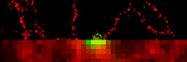
The researchers focused on determining the origin and formation of a structure only seen at the very start of the autophagy process but which gives rise to the main structure (autophagosome; the cellular 'bubble wrap') that envelops the content targeted for degradation. Due to its short-lived nature, this transient structure was difficult to characterise. The researchers jointly developed a new comprehensive imaging-based approach for observing autophagy-related structures. At the Babraham Institute this was achieved using live imaging followed by dStorm (direct Stochastic Optical Reconstruction Microscopy). At the Francis Crick Institute in London and the Zeiss Microscopy Labs in Munich, the researchers used a method called FIB-SEM (Focused Ion Beam Scanning Electron Microscopy). By combining the information gathered from these two methods, the researchers were able to identify how the first autophagy structure forms and clarify the protein and membrane associations leading to its development into a fully-fledged autophagosome.

Dr Nicholas Ktistakis, group leader in the Signalling research programme at the Babraham Institute and lead senior author, said: "By combining live imaging with cutting-edge super resolution microscopy techniques, we have been able to characterise the site of autophagy initiation and observe the physical and functional interactions between the proteins involved in autophagy. This has uncovered a new level of detail of the earliest stages of autophagy and provides a general protocol for this type of analysis in other areas of cell biology.

"Knowing more about this process increases our ability to find ways to manipulate or boost it for future therapeutic benefit."

###

This work was supported by the Biotechnology and Biological Sciences Research Council.

Media Contact

Louisa Wood
[email protected]
01-223-496-230
@babrahaminst

http://www.babraham.ac.uk/

Share12Tweet7Share2ShareShareShare1

Related Posts

Decoding Prostate Cancer Origins via snFLARE-seq, mxFRIZNGRND

February 7, 2026

Digital Health Perspectives from Baltic Sea Experts

February 7, 2026

Florida Cane Toad: Complex Spread and Selective Evolution

February 7, 2026

Exploring Decision-Making in Dementia Caregivers’ Mobility

February 7, 2026
Please login to join discussion

POPULAR NEWS

  • Robotic Ureteral Reconstruction: A Novel Approach

    Robotic Ureteral Reconstruction: A Novel Approach

    82 shares
    Share 33 Tweet 21
  • Digital Privacy: Health Data Control in Incarceration

    63 shares
    Share 25 Tweet 16
  • Study Reveals Lipid Accumulation in ME/CFS Cells

    57 shares
    Share 23 Tweet 14
  • Breakthrough in RNA Research Accelerates Medical Innovations Timeline

    53 shares
    Share 21 Tweet 13

About

We bring you the latest biotechnology news from best research centers and universities around the world. Check our website.

Follow us

Recent News

Decoding Prostate Cancer Origins via snFLARE-seq, mxFRIZNGRND

Digital Health Perspectives from Baltic Sea Experts

Florida Cane Toad: Complex Spread and Selective Evolution

Subscribe to Blog via Email

Enter your email address to subscribe to this blog and receive notifications of new posts by email.

Join 73 other subscribers
  • Contact Us

Bioengineer.org © Copyright 2023 All Rights Reserved.

Welcome Back!

Login to your account below

Forgotten Password?

Retrieve your password

Please enter your username or email address to reset your password.

Log In
No Result
View All Result
  • Homepages
    • Home Page 1
    • Home Page 2
  • News
  • National
  • Business
  • Health
  • Lifestyle
  • Science

Bioengineer.org © Copyright 2023 All Rights Reserved.