In an observational study examining bedside neuromonitoring tools in an intensive care setting, researchers found that two automated pupillometry devices yielded significantly correlated raw measurements but produced divergent clinical index scores for a substantial proportion of patients. Conducted at the Neurocritical Care Unit of San Martino Policlinic Hospital in Genoa, Italy, the study evaluated adult patients admitted with acute brain injury to determine whether the Neurologic Pupillary Index, or NPi, and the Quantitative Pupillary Index, or QPI, can be used interchangeably in clinical practice. Although the devices demonstrated strong statistical correlations when measuring basic physical pupil diameter and percentage constriction, their synthesized numerical indices frequently disagreed when categorized against established thresholds for normal and abnormal pupillary reactivity, leading investigators to conclude that the indices should not be treated as clinically interchangeable.
Assessment of the pupillary light reflex has long represented a core element of standard bedside neurological examination in patients suffering from known or suspected brain damage. Traditionally, clinicians have evaluated pupillary reactivity using manual hand-held light sources such as penlights. However, this conventional approach provides only a subjective, non-standardised qualitative measurement that is prone to considerable imprecision, driven by significant inter-observer variability and procedural heterogeneity across clinical examiners. In response to these limitations, automated pupillometry was introduced into critical care medicine to provide an objective, standardised, and highly reproducible assessment of the pupillary light reflex alongside other dynamic physiological variables, including baseline pupil size, constriction latency, constriction velocity, and dilation velocity.
Over recent years, automated pupillometry has gained wide acceptance, with the Neurologic Pupillary Index emerging as a validated metric for monitoring neurocritically ill patients. Derived from the NeurOptics NPi-300 pupillometer, the NPi is computed through a proprietary algorithm that compares multiple dynamic features of the pupillary response against a reference distribution of healthy subjects, scaling the result from 0 to 5, where values below 3 indicate abnormal pupillary light reactivity. More recently, another commercial pupillometer, the NeuroLight device developed by ID-Med, introduced the Quantitative Pupillary Index. The QPI evaluates the pupillary constriction response following a calibrated light stimulus, outputting an integer score from 0 to 5, where scores of 4 or higher define good reactivity and scores of 2 or lower denote abnormal or weak responses. Despite the increasing availability of both commercial platforms, empirical data directly comparing the performance and agreement of their respective indices in acute brain injury have remained scarce.
To investigate the clinical agreement and potential interchangeability of these two systems, the research team conducted a single-centre prospective agreement study between July 1, 2025, and December 1, 2025. The study protocol enrolled adult patients admitted to the neurocritical care unit following acute brain injury, including diagnoses such as traumatic brain injury, subarachnoid haemorrhage, intracranial haemorrhage, central nervous system infections, and intracerebral tumours. Patients presenting with severe facial or ocular trauma that precluded pupillometry, as well as those with preexisting ophthalmic diseases, were excluded from participation. The investigation was formally structured as a method-comparison and agreement study, adhering to the Guidelines for Reporting Reliability and Agreement Studies and the Strengthening the Reporting of Observational Studies in Epidemiology recommendations.
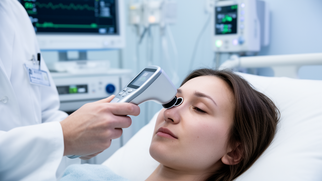
The study protocol mandated that pupillary reactivity be evaluated in both eyes of each participant using both the NeurOptics NPi-300 and the NeuroLight pupillometer. The instruments were applied in a random order without predefined criteria, separated by a 60-second recovery interval between measurements to allow the pupil sufficient time to return to its baseline dimension following initial light stimulation. To avoid acute confounding, clinical care teams maintained constant mechanical ventilation and sedation regimens during the paired recording sessions, and only the first combined measurement session conducted within the first seven days of admission was included in the analysis. The hardware specifications differed slightly between devices: the NPi-300 utilized an infrared camera with fixed light stimulation of 1000 lux for 3.2 seconds, while the NeuroLight generated a light stimulus of 320 lux for 1 second, capturing pupil diameter and constriction within 4 seconds of stimulation onset.
A total of 76 patients were initially screened during the study period, of whom 3 were excluded due to concurrent ocular trauma, resulting in a final analytical cohort of 73 patients. The median age of the cohort was 65 years, with an interquartile range spanning 54 to 77 years, and 45 patients, representing 62 percent of the group, were male. The underlying clinical conditions precipitating intensive care admission included traumatic brain injury in 26.0 percent of cases, haemorrhagic stroke in 19.2 percent, and subarachnoid haemorrhage and ischemic stroke in 15.1 percent each. At the time of pupillary assessment, 89.0 percent of patients were undergoing mechanical ventilation and 42.5 percent were receiving vasopressor support. Analgesic and sedative medications were actively administered to 84.9 percent of the cohort, including fentanyl in 79.5 percent, propofol in 56.2 percent, dexmedetomidine in 9.6 percent, and midazolam in 5.5 percent.
Primary agreement between the devices was evaluated using Bland-Altman analysis to quantify systematic bias and limits of agreement, supported by secondary Spearman rank correlation coefficients. The raw physiological metrics showed strong alignment across devices: the mean bias for pupil diameter was 0.44 millimetres, with 95 percent limits of agreement ranging from minus 0.43 to 1.31 millimetres, while pupillary reactivity demonstrated a mean bias of 0.57 percent, with limits of agreement between minus 7.63 percent and 8.77 percent. Spearman correlation coefficients indicated very strong associations for both baseline pupil diameter, at rho equals 0.91, and pupillary constriction percentage, at rho equals 0.92, with both achieving statistical significance at p values below 0.0001.
However, when comparing the derived indices directly, Bland-Altman analysis revealed a systematic mean bias of minus 0.82 between NPi and QPI, accompanied by wide limits of agreement spanning from minus 2.70 to 1.05 units. Although Spearman rank correlation remained statistically robust at rho equals 0.75, the clinical agreement between index categories demonstrated notable discordance. Across the cohort, 30 patients, or 40.1 percent, exhibited an abnormal pupillary response on at least one device based on established normality cut-offs. Overall, inter-device classification discordance occurred in 21 patients, representing 28.8 percent of paired measurements. Among these discordant cases, 16 patients showed an abnormal QPI alongside a normal NPi, whereas 5 patients exhibited an abnormal NPi despite a normal QPI, yielding a Cohen’s kappa coefficient of 0.28, which corresponds to only fair agreement beyond chance.
The evaluation of anisocoria, defined as a difference in pupillary diameter between eyes of 0.5 millimetres or greater, also revealed moderate discrepancies between the two platforms. The NeuroLight pupillometer identified anisocoria in 18 patients, representing 24.7 percent of the cohort, while the NPi-300 identified 19 cases, representing 26.0 percent. When comparing paired determinations across patients, the two devices disagreed in 15 cases, representing 20.5 percent of the sample, which yielded a Cohen’s kappa coefficient of 0.47, indicating only moderate inter-device agreement for detecting pupillary asymmetry at the bedside.
The authors observed that the apparent divergence between strong correlation and poor clinical interchangeability stems from methodological and algorithmic differences in how the two indices are calculated. The NPi relies on a multidimensional, composite calculation that incorporates multiple dynamic kinematic features—including constriction velocity, latency, amplitude, and dilation velocity—each normalized against a normative reference population, which may confer stability against isolated physiological perturbations. In contrast, the QPI is predominantly driven by pupillary constriction velocity and percentage constriction, rendering it potentially more sensitive to external influences such as ambient lighting conditions, baseline diameter, and pharmacological depression from sedatives and opioids. Because sedatives like propofol and opioids frequently blunt pupillary constriction kinetics in intensive care populations, single-parameter weighted metrics may register abnormal values where composite scores remain buffered.
The authors highlighted several methodological limitations that temper the interpretation of their findings. The investigation was conducted at a single centre with a relatively modest sample size of 73 patients, and it utilized an exploratory agreement design that was not powered to assess prognostic accuracy or long-term clinical outcomes. Furthermore, pupillary evaluations were recorded across a broad clinical window of up to seven days following intensive care admission, during which ongoing medical interventions, sedation depth, and intracranial dynamics varied naturally. The study did not incorporate continuous electroencephalography, Bispectral Index, or Nociception Level monitoring to standardize sedation and nociception objectively during measurements, nor did it track granular drug dosing schedules. Additionally, the NeuroLight pupillometer utilizes an ambient-light-excluding silicone eyecup, introducing physical acquisition differences compared to the NPi-300.
In their final assessment, the researchers emphasized that while automated pupillometry devices reliably record core physiological variables like pupil size, clinicians must exercise caution when interpreting derived scores across different hardware platforms. Because the NPi and QPI can classify more than a quarter of patients into conflicting clinical categories around standard diagnostic thresholds, the two indices cannot be assumed to provide equivalent bedside information. The investigators concluded that further large-scale, prospective multicentre studies will be necessary to establish the distinct clinical roles, diagnostic utility, and comparative prognostic significance of each pupillometric index in patients with acute brain injury.
Subject of Research: Comparison of Neurologic Pupillary Index and Quantitative Pupillary Index in neurocritical care
Article Title: A comparison between two indices derived from automated pupillometry devices
Article References: Bandoni, C., Mounir, K., Iannuzzi, F., Berardino, A., Tarantino, F., Andaloussi, M. R., Taccone, F. S., Battaglini, D., Patroniti, N. A., & Robba, C. (2026). A comparison between two indices derived from automated pupillometry devices. Journal of Clinical Monitoring and Computing. https://doi.org/10.1007/s10877-026-01490-4
Image Credits: AI Generated
DOI: 10.1007/s10877-026-01490-4
Keywords: automated pupillometry, neurologic pupillary index, quantitative pupillary index, neurocritical care, acute brain injury, pupillary light reflex, Bland-Altman analysis, clinical monitoring, traumatic brain injury, intensive care unit, inter-device agreement, anisocoria, pupil diameter, pupillary constriction, Neuroptics NPi-300, NeuroLight pupillometer
News Source: Cassandra Pierce. (November 20, 2026). Automated Pupillometry Indices Differ Significantly in Evaluating Critically Ill Patients. Scienmag.