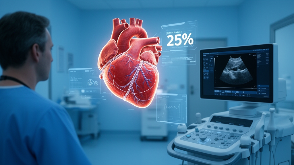
A fully automated artificial intelligence system evaluated in a prospective pilot study completed comprehensive transthoracic echocardiography analyses in a median time of 94 seconds per examination compared with 490 seconds required by a trained human sonographer, while achieving noninferior diagnostic accuracy for left ventricular ejection fraction against an expert cardiologist reference standard. Published in the Journal of Cardiovascular Imaging, the investigation examined whether integrating automated quantitative measurement algorithms with automated qualitative visual interpretation modules could alleviate the operational burden imposed by escalating imaging volumes in clinical cardiology laboratories without compromising diagnostic fidelity. In routine cardiovascular care, comprehensive transthoracic echocardiography requires clinicians and sonographers to integrate numerous manual quantitative measurements, spanning chamber dimensions, volumes, and blood flow velocities, with visual qualitative interpretations of myocardial wall motion, pericardial abnormalities, and valvular pathologies. Because conventional manual analyses remain inherently operator-dependent and resource-intensive, previous automated attempts often addressed isolated quantitative tasks rather than combining quantitative calculations with visual interpretation, leaving end-to-end clinical workflow efficiency largely unaddressed under standardized conditions.
To evaluate the real-world feasibility of end-to-end automation, researchers at Seoul National University Bundang Hospital assembled an exploratory cohort of forty transthoracic echocardiography examinations obtained from adult patients between November and December 2025 under Institutional Review Board approval. The study design employed a prospective, paired comparison protocol using identical, deidentified Digital Imaging and Communications in Medicine datasets provided independently to both a human analyst and an automated software pipeline. Eligible examinations were required to fulfill a rigorous core acquisition checklist comprising standard B-mode views, including parasternal long-axis, parasternal short-axis at the great vessels, mitral valve, and papillary muscle levels, a right ventricular-focused view, and apical four-chamber, two-chamber, and three-chamber views. In addition, qualifying datasets included M-mode recordings of the aortic root, left atrium, and left ventricle, alongside spectral Doppler acquisitions covering continuous-wave Doppler across the aortic valve, pulsed-wave Doppler of the left ventricular outflow tract, continuous-wave Doppler of tricuspid regurgitation, and septal tissue Doppler imaging.
The study protocol applied strict exclusion criteria to eliminate confounding factors, potential data leakage, and unmeasurable imaging artifacts. The investigators excluded repeat examinations from identical patients during the screening window and discarded examinations missing fundamental demographic or anthropometric metadata, such as age, sex, height, or weight. Furthermore, examinations were excluded if image quality was determined to be unmeasurable to the extent that expert adjudication of core measurements or qualitative assessments could not be established. To prevent artificial performance inflation resulting from training data leakage, the researchers explicitly excluded patients known to have been registered in the Open AI Dataset Project supported by the Korean Ministry of Science and ICT, as the evaluated commercial software had incorporated data from that national repository during its development. For eligible datasets, all previous measurement annotations, graphical overlays, and derived report texts were stripped from the digital files before anonymized files were assigned unique study codes for parallel distribution.
The automated software platform evaluated in the trial comprised two specialized functional components developed by Ontact Health: a quantitative measurement module designated SoniX EchoQuant and an investigational qualitative visual interpretation module designated SoniX EchoVerse. While SoniX EchoQuant was commercially available as part of SONIX Health version 2.0 and already operating routinely at the host institution, SoniX EchoVerse was deployed experimentally during the study while undergoing regulatory review. The investigative team incorporated the unapproved visual interpretation module specifically to establish an equitable operational comparison against human practice, noting that sonographers simultaneously evaluate qualitative visual findings while measuring quantitative indices, meaning an evaluation restricted solely to quantitative automation would underestimate human work scope. Both algorithmic components functioned autonomously without operator intervention, executing automatic view recognition and producing up to seventy-five quantitative indices and one hundred sixty-two qualitative assessment items when required acoustic views were available.
The computational pipeline executed within a dedicated local computing environment equipped with a forty-eight-core Intel Xeon w7-3455 central processing unit and an NVIDIA RTX A6000 graphics processing unit. For the human comparison arm, a single trained cardiac sonographer with six years of clinical experience performed independent manual quantitative measurements and visual assessments using the hospital’s clinical INFINITT Cardiology PACS workstation. The human reader remained completely blinded to original clinical reports and all artificial intelligence outputs throughout the protocol. Human analysis duration was captured objectively by logging the elapsed time between the initial loading of a digital examination into the picture archiving software and the formal completion of a standardized core checklist matching the institution’s clinical reporting structure. To establish the ground-truth reference standard, an experienced board-certified cardiologist possessing more than fifteen years of echocardiographic experience independently adjudicated the complete image series, re-measuring quantitative parameters and grading qualitative findings under strict blinded conditions.
The study cohort encompassed diverse clinical presentations across forty adult examinations, reflecting purposeful sampling designed to evaluate algorithmic stability across varying cardiac rhythms, image clarities, and functional states. The median patient age was 73.5 years, with an interquartile range of 62.0 to 83.0 years, and twelve patients, representing 30.0 percent of the cohort, were male. The median body surface area measured 1.69 square meters, with an interquartile range of 1.56 to 1.85 square meters. Sinus rhythm was documented in twenty-seven examinations, or 67.5 percent, while thirteen examinations, representing 32.5 percent, presented with atrial fibrillation. Subjective acoustic image quality among included studies was rated as good in nineteen cases, average in seventeen cases, and poor in four cases. Left ventricular systolic function varied across the cohort: twenty-eight examinations showed an ejection fraction exceeding 55 percent, nine examinations ranged between 35 and 55 percent, and three examinations demonstrated severe systolic dysfunction with values below 35 percent.
Statistical evaluation of workflow duration demonstrated a significant efficiency advantage for the automated computational pipeline. Across all forty examinations, both the human reader and the dual-module artificial intelligence platform achieved a 100 percent processing completion rate without analytical failures. The human workflow exhibited a median completion time of 490 seconds, with an interquartile range spanning 438 to 626 seconds and total durations ranging from 370 to 1,127 seconds. In contrast, the fully automated artificial intelligence workflow achieved a median total processing time of 94 seconds, with an interquartile range of 82 to 106 seconds and a range of 69 to 121 seconds. Component runtime tracking showed that the SoniX EchoQuant module consumed a median of 74 seconds with an interquartile range of 68 to 83 seconds, while the SoniX EchoVerse visual module required a median of 18 seconds with an interquartile range of 15 to 22 seconds. Paired comparisons conducted via the Wilcoxon signed-rank test confirmed that the total workflow duration reduction was statistically significant, yielding a P-value of less than 0.001.
Evaluation of the prespecified primary accuracy endpoint confirmed that the artificial intelligence system achieved noninferior estimation of Simpson biplane left ventricular ejection fraction when evaluated against the adjudicated reference standard. Working with a predefined noninferiority margin of 3 percentage points at a one-sided significance level of 0.025, the mean difference between algorithmic estimates and expert reference values was 0.00 percentage points with a standard deviation of 5.29 percentage points. The upper bound of the one-sided 95 percent confidence interval reached 1.41 percentage points, remaining well beneath the 3 percentage point margin and confirming noninferiority with a P-value of less than 0.001. Absolute agreement analysis yielded an intraclass correlation coefficient of 0.902 with a 95 percent confidence interval of 0.822 to 0.947, while Bland-Altman analysis demonstrated zero mean systematic bias, accompanied by 95 percent limits of agreement extending from minus 10.37 to positive 10.37 percentage points. The mean absolute error for algorithmic ejection fraction was 3.53 percentage points, with a median absolute error of 2.53 percentage points.
Benchmarking comparisons involving the human analyst revealed distinct operational characteristics relative to the cardiologist reference standard. Unlike the artificial intelligence system, which displayed no systematic directional bias, human sonographer estimates demonstrated a mild tendency toward overestimation, with a mean positive difference of 2.95 percentage points, a 95 percent confidence interval from 1.26 to 4.64, and a standard deviation of 5.29 percentage points. The human intraclass correlation coefficient for left ventricular ejection fraction was 0.88 with a 95 percent confidence interval of 0.71 to 0.94. The human reader produced a mean absolute error of 4.67 percentage points and a median absolute error of 3.43 percentage points. While investigators noted that comparisons involving a single reader serve as supportive descriptive benchmarking rather than evidence of broader human interobserver tendencies, the finding underscored the algorithmic platform’s ability to maintain stable estimation of global left ventricular systolic performance across standardized inputs.
Secondary quantitative indices produced by the automated system showed generally robust agreement with reference measurements across thirty-nine or forty paired examinations, yielding intraclass correlation coefficients spanning from 0.625 to 0.989. Agreement was highest among Doppler blood flow velocities and chamber volume calculations; continuous-wave spectral Doppler peak early diastolic filling velocity achieved an intraclass correlation coefficient of 0.989, left ventricular end-systolic volume achieved 0.950, left ventricular end-diastolic volume achieved 0.940, and left atrial volume reached 0.9333. Conversely, lower correlation coefficients were documented for myocardial wall-thickness measurements, including the interventricular septum and posterior left ventricular wall. Investigators noted that despite lower correlation coefficients, the median absolute errors and mean differences for wall thicknesses remained close to 1 millimeter, suggesting that restricted physiological measurement ranges and heightened sensitivity to border demarcation or beam orientation contributed to lower statistical correlations despite modest absolute differences.
Assessment of the secondary qualitative endpoint revealed moderate to substantial agreement in grading aortic regurgitation severity across forty examinations enriched with valvular lesions. Reference adjudication classified nineteen examinations as none or trivial, ten as mild, eight as moderate, and three as severe. The artificial intelligence platform attained an overall exact-grade accuracy of 75.0 percent and a quadratic weighted kappa of 0.762. However, class-specific sensitivity varied substantially across severity tiers: the algorithm achieved a sensitivity of 1.00 for none or trivial, 0.90 for mild, 0.00 for moderate, and 0.67 for severe regurgitation, culminating in a balanced accuracy of 0.642. For comparison, human interpretation achieved an overall accuracy of 82.5 percent, a quadratic weighted kappa of 0.812, and a balanced accuracy of 0.824, with sensitivities of 0.947 for none or trivial, 0.60 for mild, 0.75 for moderate, and 1.00 for severe disease. In paired exact-grade accuracy assessments, both modalities agreed with the reference standard in twenty-five examinations, both erred in two, the algorithm alone was correct in five, and the human reader alone was correct in eight, yielding no statistically significant difference under McNemar testing with a P-value of 0.579.
The study authors highlighted several methodological limitations that contextualize their findings and define avenues for subsequent research. Because the investigation was conducted as an exploratory, single-center pilot trial comprising forty purposively sampled examinations, the observed effect sizes serve as hypothesis-generating metrics rather than definitive population-level performance benchmarks. Furthermore, the human comparison arm relied on a single sonographer and a single adjudicating cardiologist, precluding broader assessments of interobserver variability among diverse clinicians. Additionally, the qualitative visual interpretation module represented an early developmental version, and detailed qualitative reporting was largely restricted to aortic regurgitation grading. Finally, because all imaging data were collected within a single tertiary medical center using local scanning protocols and executed on a specific hardware configuration, multicenter trials across diverse ultrasound machine vendors, clinical staffing models, and hospital networks will be required before automated pipelines can be widely integrated into routine clinical reporting workflows.
Subject of Research: Automated artificial intelligence workflow efficiency and measurement accuracy in transthoracic echocardiography
Article Title: Fully automated artificial intelligence–based echocardiographic analysis substantially reduces workflow time while preserving measurement accuracy: a pilot study
Article References: Sun, J., Yoon, Y. E., Lee, J., Lee, G., Bak, M., Park, J., Choi, H.-M., Hwang, I.-C., & Cho, G.-Y. (2026). Fully automated artificial intelligence–based echocardiographic analysis substantially reduces workflow time while preserving measurement accuracy: a pilot study. Journal of Cardiovascular Imaging, 34(1), Article 10. https://doi.org/10.1186/s44348-026-00073-w
Image Credits: AI Generated
DOI: 10.1186/s44348-026-00073-w
Keywords: transthoracic echocardiography, artificial intelligence, left ventricular ejection fraction, workflow efficiency, SoniX EchoQuant, SoniX EchoVerse, aortic regurgitation, intraclass correlation coefficient, cardiovascular imaging, DICOM datasets, noninferiority trial, cardiac sonography, Bland-Altman analysis, Doppler echocardiography, clinical PACS, cardiac chamber volume
News Source: Blake Davidson. (November 19, 2026). Automated Echocardiography Artificial Intelligence Cuts Analysis Time While Preserving Ejection Fraction Accuracy. Scienmag.