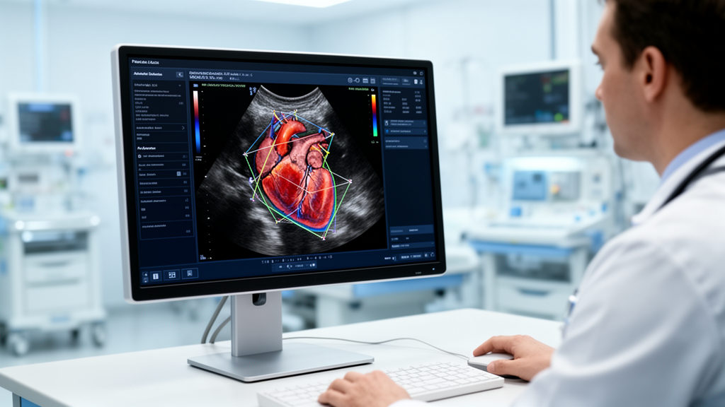
Echocardiography remains the foundational imaging modality across cardiovascular medicine, prized by clinicians for its portability, real-time diagnostic capabilities, and favorable safety profile. Yet conventional transthoracic echocardiography workflows—requiring manual probe manipulation and image acquisition by specialized sonographers, sequential caliper tracing of structural and functional parameters, and comprehensive clinical interpretation by cardiologists—face severe operational bottlenecks. Escalating patient volumes, persistent specialist workforce shortages, and the physical toll of repetitive transducer manipulation have strained hospital laboratories. Diagnostic throughput is further constrained by the manual measurement phase, which demands the extraction of dozens of individual parameters tailored to complex disease presentations while introducing substantial interobserver variability that can compromise longitudinal clinical consistency. To resolve these compounding systemic pressures, medical researchers have accelerated the deployment of artificial intelligence systems, transitioning computational tools from exploratory proof-of-concept models to prospective clinical evaluations designed to automate quantitative tasks and restructure diagnostic pathways.
The structural integration of machine learning into clinical echocardiography follows a defined spectrum of autonomy, ranging from assistive systems that support operator guidance to end-to-end automated pipelines. Assistive tools directly support sonographers during examination sessions through automated view recognition, image optimization, and real-time measurement suggestions. In contrast, integrated workflows embed algorithmic processing across multiple contiguous stages of scanning, quantitative analysis, and preliminary report drafting. Fully automated architectures generate discrete task-specific outputs without human manual manipulation, whereas human-in-the-loop governance models enforce mandatory clinician verification, adjustment, and authorization before diagnostic conclusions enter permanent medical records. Across this spectrum, modern algorithmic implementations increasingly diverge from opaque, black-box pattern recognition frameworks. Instead, emerging architectures formalize expert clinical reasoning, combining anatomical visualization with hemodynamic measurements to establish inspectable, multi-parameter diagnostic assessments.
Substantial empirical evidence demonstrates that algorithmic automation directly alleviates the physical and ergonomic strains inherent to high-volume echocardiographic scanning. Sonographers frequently suffer from work-related musculoskeletal disorders caused by sustained transducer pressure, awkward upper-body posture, and repetitive console interactions. In an ergonomic evaluation conducted by Hollitt and colleagues, the deployment of an artificial intelligence protocol incorporating automatic view classification and guided measurement caliper placement reduced console keystroke volume from 503 to 231 actions per study. Concurrently, active scanning duration fell from 18.0 minutes to 13.4 minutes without degrading image diagnostic quality. Subsequent investigations demonstrated that unifying cardiac cycle phase selection, view detection, and left ventricular caliper placement into single-step automated routines produces even larger ergonomic improvements, confirming that computational integration mitigates the cumulative physical burdens driving occupational injury among imaging professionals.
Beyond physical ergonomics, prospective clinical investigations demonstrate measurable enhancements in institutional workflow efficiency, scanning throughput, and parameter completeness. In a randomized crossover trial conducted in Japan across 38 dedicated study days and 585 screening examinations, researchers evaluated an artificial intelligence-assisted protocol against conventional manual workflows, with attending cardiologists reviewing and finalizing all clinical reports while blinded to study arm assignments. The computational protocol achieved a modest reduction in examination duration from 14.3 ± 4.2 minutes down to 13.0 ± 3.5 minutes (P < 0.001) while expanding the average number of quantified parameters per examination by 3.4-fold (P < 0.001). This diagnostic expansion lifted overall laboratory throughput from 14.1 ± 2.5 examinations per day to 16.7 ± 2.5 examinations per day (P = 0.003) and raised objective image quality scores (P < 0.001). Concurrently, sonographer fatigue scores decreased from 4.7 ± 0.6 in the standard arm to 4.1 ± 1.1 under assisted scanning (P = 0.039), while maintaining a 90 percent concordance rate with expert-adjudicated parameter thresholds. Demonstrating similar efficiency gains, an independent automated quantification platform compressed downstream analytical processing times from 490 seconds to 94 seconds.
Rigorous clinical trials have validated the quantitative precision of deep learning models across routine ventricular functional assessments. In a blinded, randomized noninferiority trial evaluating left ventricular ejection fraction quantification, initial measurements required substantial clinical modification—defined as an absolute deviation greater than 5 percent upon expert cardiologist review—in only 16.8 percent of algorithm-assisted examinations compared with 27.2 percent of conventional sonographer-measured examinations. Reviewing cardiologists could not reliably differentiate machine-derived contour determinations from manual sonographer tracings, and the automated workflow generated significant time savings across both sonography and cardiology teams. In a complementary pilot study encompassing 40 transthoracic echocardiograms that deliberately included challenging acoustic windows, automated ejection fraction estimates matched expert adjudication with an intraclass correlation coefficient of 0.90. Extending these findings across broader diagnostic sets, a formal validation study of 602 echocardiograms evaluated 23 discrete anatomical parameters; deep learning measurements showed less divergence from any individual human expert than the three blinded human readers demonstrated among themselves, meeting prespecified noninferiority criteria for every evaluated parameter.
To overcome algorithmic discrepancies stemming from proprietary architectures, investigators have prioritized open-source platforms validated across diverse healthcare environments. Sahashi and colleagues developed an open-source deep learning framework capable of quantifying 18 distinct anatomical and Doppler parameters. Initially trained on extensive clinical cohorts at Cedars-Sinai Medical Center, the model underwent external validation at Stanford Healthcare, sustaining robust diagnostic performance across patient subgroups categorized by age, sex, body mass index, atrial fibrillation status, and ultrasound equipment manufacturer. Transparent, publicly accessible frameworks provide reproducible scientific benchmarks that counter the opacity of proprietary commercial systems, establishing standardized baselines for quantitative reliability across heterogeneous clinical populations.
A critical clinical advantage of automated imaging platforms lies in their ability to capture comprehensive diagnostic markers that are routinely omitted during manual workflows due to time constraints. International clinical guidelines—including the 2025 American Society of Echocardiography recommendations on diastolic function, joint guidance from the European Association of Cardiovascular Imaging, and British Society of Echocardiography guidelines—have established left atrial reservoir strain and global longitudinal strain as essential metrics for grading diastolic dysfunction and monitoring cardiotoxicity. However, conventional speckle-tracking requires specialized post-processing software and cumbersome manual contour adjustments. In a retrospective cohort study of 731 hospitalized patients, baseline mortality prediction models relying on standard manual parameters exhibited comparable predictive performance between artificial intelligence and human experts, yielding receiver operating characteristic areas under the curve of 0.67 and 0.66 (P = 0.86). When automated global longitudinal strain—routinely captured by the algorithm but omitted during manual clinical examinations—was incorporated into the model, predictive accuracy improved significantly to an area under the curve of 0.73 (P = 0.048). Notably, 18 percent of patient studies were excluded from this analysis, including approximately 8 percent rejected due to poor acoustic windows, highlighting ongoing technical barriers in fragile clinical cohorts.
Recent computational developments have moved beyond isolated parameter estimation toward multi-task networks capable of comprehensive study interpretation. Holste and colleagues trained a multi-task deep learning model on 1.2 million echocardiographic video sequences derived from 32,265 clinical studies at Yale New Haven Health System, engineering the system to evaluate 18 diagnostic classification tasks alongside 21 continuous parameter measurements in a unified inference step. The architecture achieved a median area under the curve of 0.91 across classification endpoints, demonstrating exceptional accuracy for moderate or worse left ventricular systolic dysfunction (areas under the curve of 0.98 internally and 0.99 externally) and severe aortic stenosis (areas under the curve of 0.98 internally and 1.00 externally), alongside mean absolute errors for ejection fraction estimation of 4.2 percent and 4.5 percent. When deployed on emergency department point-of-care ultrasound recordings, the model maintained a median area under the curve of 0.85 across 14 diagnostic targets. However, predicting critical structural pathologies directly from pixel data without verifiable intermediate Doppler envelopes creates interpretability challenges that can hinder clinical adoption.
To overcome the limitations of uninterpretable pixel classifiers, modern diagnostic architectures synthesize structural video with quantitative hemodynamics to emulate specialist clinical reasoning. In valvular disease, Park and colleagues developed an artificial intelligence framework for aortic stenosis grading that couples structural cardiac imaging with quantitative continuous-wave Doppler velocity pattern analysis, enhancing both initial diagnostic categorization and long-term prognostic modeling. In myocardial infiltration, computational texture analysis formalizes the subjective visual feature known as granular sparkling; algorithms extract pixel-level spatial features using gray-level co-occurrence matrices and neighborhood gray-tone difference matrices to identify cardiac amyloidosis. Advanced models integrate direct pixel intensity assessments to distinguish transthyretin amyloid cardiomyopathy from light-chain amyloidosis and clinical phenocopies, with multi-view implementations leveraging Shapley Additive Explanations to expose decision drivers. Similarly, in pericardial disease, Chao and colleagues established deep learning differentiation of constrictive pericarditis from restrictive cardiomyopathy on apical four-chamber views to expedite surgical referral. Extending this logic, Jeong and colleagues introduced a two-stage sequential reasoning framework that first assesses pericardial effusion volume and thickening on B-mode views before integrating Doppler spectra and inferior vena cava dimensions to determine hemodynamic significance.
Beyond central hospital laboratories, machine learning models are expanding access to cardiovascular assessments across point-of-care, ambulatory, and underserved environments. Narang and colleagues demonstrated that registered nurses without prior ultrasound experience could acquire diagnostic-quality transthoracic views using real-time automated navigation tools after brief instruction. In outpatient heart failure pathways, a prospective multicenter evaluation of 587 patients across five clinics evaluated handheld automated echocardiography without excluding degraded recordings. The automated device identified left ventricular ejection fractions of 40 percent or less with an area under the curve of 0.96, specificity of 0.95, and negative predictive value of 0.97, establishing its utility as an outpatient triage tool, although a modest sensitivity of 0.61 demonstrated that suboptimal acoustic quality can cause reduced functional capacity to go undetected. In oncology clinics, standardized training enabled oncologists and nurses to track ejection fractions in 115 patients with accuracy comparable to expert sonography. Complementing these clinical expansions, health economic modeling in Singapore demonstrated institutional cost-effectiveness through specialist task-shifting, while tele-echocardiography frameworks described by Kusunose offer scalable imaging support for geographically isolated medical centers.
Despite documented operational gains, critical methodological and governance challenges preclude uncritical clinical implementation. Much of the supporting literature relies on small cohorts and single-center academic designs; the Japanese randomized trial enrolled four sonographers, and landmark ergonomic and language-model studies examined restricted operational settings. Furthermore, algorithm accuracy remains uneven across cardiac pathologies: left ventricular ejection fraction is rigorously validated, whereas right ventricular quantification, multi-parametric diastolic grading, and complex valvular lesions exhibit variable fidelity. Intervendor variability represents an additional hurdle, as Szasz and colleagues demonstrated that different commercial algorithms generate discordant measurements on identical imaging datasets. Image degradation remains an operational bottleneck, evidenced by the exclusion of approximately 7 percent of studies in blinded ejection fraction trials and 8 percent in automated strain cohorts due to inadequate acoustic penetration. Concurrently, utilizing large language models to draft unstructured diagnostic reports introduces risks of automation bias, where overburdened clinicians passively endorse incorrect machine-generated text. Because regulatory frameworks treat automated diagnostic algorithms as Software as a Medical Device while assigning legal liability exclusively to the signing cardiologist, clinical facilities must establish robust governance protocols, require verifiable visual intermediate outputs, and embed formal artificial intelligence literacy into cardiovascular training curricula.
Subject of Research: Artificial intelligence integration and clinical reasoning in echocardiography workflows
Article Title: Reinventing the echocardiography workflow: from manual quantification to artificial intelligence–driven comprehensive interpretation
Article References: Heo, R., Lee, S.-A., & Chang, H.-J. (2026). Reinventing the echocardiography workflow: from manual quantification to artificial intelligence–driven comprehensive interpretation. Journal of Cardiovascular Imaging, 34(1), Article 27. https://doi.org/10.1186/s44348-026-00087-4
Image Credits: AI Generated
DOI: 10.1186/s44348-026-00087-4
Keywords: echocardiography, artificial intelligence, cardiovascular imaging, left ventricular ejection fraction, myocardial strain, workflow efficiency, sonographer ergonomics, deep learning, point-of-care ultrasound, clinical decision support, valvular heart disease, pericardial disease, cardiac amyloidosis, diagnostic reasoning, automation bias, intervendor variability, handheld ultrasound
News Source: Ophelia Keating. (November 18, 2026). Artificial Intelligence Reshapes Echocardiography Workflows and Diagnostic Reasoning Across Clinical Practice. Scienmag.