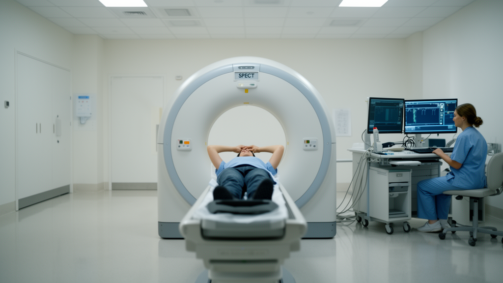
A cardiac phantom study has found that the choice of acquisition mode and the position of a patient’s arms can meaningfully change the quality of myocardial perfusion images obtained with a full-ring semiconductor SPECT/CT system. The research, published in EJNMMI Physics, used an electrocardiography-gated myocardial phantom rather than human patients, and its conclusions therefore describe scanner performance under controlled laboratory conditions rather than confirmed clinical outcomes.
Full-ring semiconductor SPECT/CT systems offer a focus mode designed to enhance image quality and shorten acquisition time by concentrating data collection on the heart, alongside a uniform mode that covers the whole body contour. The study’s authors noted that the impact of arm positioning on focus mode had remained under-investigated, prompting a systematic comparison of both modes under two arm configurations.
The team built the experiment around an ECG-gated myocardial phantom filled with Tc-99m solution adjusted to simulate the radiotracer concentration found in the myocardium. To mimic the arms-down position, they attached the upper limb portion of a whole-body phantom; removing it simulated the arms-up position. Acquisitions were then performed in both arm positions using the uniform mode and the focus mode, with contrast analysis, polar map assessment and cardiac functional analysis carried out for every condition.
The count measurements revealed a clear trade-off between the two modes. Under all conditions, the number of myocardial counts was higher in uniform mode than in focus mode, but the number of ventricular cavity counts was significantly reduced in focus mode.
When CT-based attenuation correction was applied, the mean myocardial count in focus mode was 11,075 ± 1064 with the arms raised and 9467 ± 843 with the arms lowered, while ventricular cavity counts fell to 261 ± 191 and 631 ± 318 respectively. As a result, focus mode consistently achieved superior contrast ratios, reaching a maximum value of 0.98 ± 0.03 in the arms-raised position when attenuation and scatter correction were combined.
Polar map analysis added a spatial dimension to the findings. Focus mode improved myocardial uniformity and mitigated apparent uptake reductions in the septal and lateral walls compared with uniform mode, an effect that was particularly pronounced under the arms-down condition.
Quantitative gated SPECT analysis showed that end-diastolic volume was underestimated across all modes, but the two modes diverged sharply on ejection fraction. Focus mode produced EF values of 27.7 percent (range 27.6–27.8) for the arms-down position and 27.1 percent (range 25.7–28.5) for arms-up, closely matching the phantom’s theoretical value of 27.7 percent. Uniform mode, by contrast, significantly overestimated EF, particularly in the arms-down condition, where it reached 35.4 percent (range 33.1–37.7).
Based on these phantom measurements, the researchers concluded that focus mode acquisition with the arms raised represents the optimal imaging approach for maximizing image contrast and supporting reliable assessment of cardiac functional parameters on full-ring semiconductor SPECT/CT systems. They also emphasized a practical secondary benefit: focus mode maintained similar quantitative accuracy even when imaging was performed with the arms down, which could enable a relatively high level of evaluation in patients with limited physical mobility.
Because the work was performed with a phantom rather than patients, the reported counts, contrast ratios and functional values reflect controlled scanner behavior under idealized conditions, and the authors’ conclusions about clinical utility will require validation in patient studies. Nevertheless, the findings offer concrete guidance for nuclear medicine laboratories operating full-ring semiconductor systems: where patient condition allows, acquiring gated myocardial perfusion images in focus mode with the arms raised appears to deliver the best combination of contrast and quantitative accuracy, while focus mode with arms down remains a strong fallback option.
Subject of Research: Effects of acquisition mode and arm position on ECG-gated myocardial perfusion SPECT imaging with a full-ring semiconductor SPECT/CT, tested in a phantom study.
Article Title: Effects of acquisition mode and arm position on myocardial perfusion imaging with a full-ring semiconductor SPECT/CT: ECG-gated myocardial phantom study
Article References: Hatano, A., Miyoshi, A., Odagiri, H., Hebiguchi, H., Oura, K., Jimbo, H., Tanaka, Y., Takeuchi, T., Kodama, H., Takanami, K., Usui, A., & Kaneta, T. (2026). Effects of acquisition mode and arm position on myocardial perfusion imaging with a full-ring semiconductor SPECT/CT: ECG-gated myocardial phantom study. EJNMMI Physics. https://doi.org/10.1186/s40658-026-00958-1
Image Credits: AI Generated
DOI: 10.1186/s40658-026-00958-1
Keywords: SPECT/CT, myocardial perfusion imaging, semiconductor detector, focus mode, arm position, cardiac phantom, ECG gating, attenuation correction, QGS, ejection fraction, nuclear cardiology, image contrast
News Source: Denise Maddox. (October 28, 2026). Arms Up and Focus Mode Deliver Sharper Heart SPECT Images in Phantom Study. Scienmag.