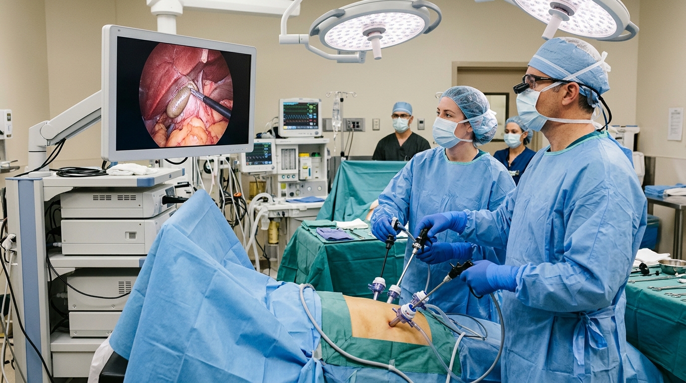
Laparoscopic cholecystectomy is widely regarded as one of the most common surgical procedures performed globally, favored for its association with faster patient recovery, reduced postoperative pain, and improved cosmetic outcomes. However, the prevalence of this surgery means that even infrequent complication rates result in a significant number of incidents. New research highlights that variations in the origin, course, number, and branching patterns of the cystic artery—the vessel supplying blood to the gallbladder—act as independent, yet often overlooked, contributors to operative complexity and the risk of vasculobiliary injury. While established surgical training emphasizes achieving a critical view of safety to prevent bile duct injuries, a recent case series suggests that surgeons must pay closer attention to complex vascular anatomy to mitigate risks during these routine operations.
This retrospective observational case series analyzed six laparoscopic cholecystectomies where a gastrointestinal surgeon encountered direct intraoperative influences from vascular anomalies. By examining these real-time challenges, the study illustrates that vascular variations are not merely anatomical curiosities but critical factors that can dictate intraoperative decision-making and patient safety. The researchers reviewed relevant medical literature to provide a comprehensive surgical perspective on managing these non-classical scenarios, identifying several distinct patterns that deviate from standard textbook configurations. The findings underscore that recognizing these variations is essential for surgeons to maintain low complication rates, particularly when the typical anatomical landmarks of Calot’s triangle are distorted or obscured.
Standard anatomical configuration typically features a single cystic artery positioned behind the common hepatic duct and anterior to the cystic duct within the boundaries of Calot’s triangle. Deviations from this standard are frequent, occurring in approximately 15 to 25 percent of cases. These low-lying arteries can lie anterior and superior to the common bile duct, carrying a heightened risk of hemorrhage due to peritoneal involvement during dissection. Furthermore, the study cataloged positional differences, such as arteries coursing posterior to the cystic duct, and noted that the anterior cystic artery is particularly prone to accidental injury because it is often the first structure a surgeon encounters.
One of the more hazardous configurations identified is the “caterpillar hump,” or Moynihan’s hump, where the right hepatic artery becomes tortuous and occupies a significant portion of Calot’s triangle. This tortuosity shortens the cystic artery and presents a severe risk of misidentification, where a surgeon might mistakenly ligate the right hepatic artery instead of the cystic artery, leading to potential necrosis of the right side of the liver. The case series also highlighted instances of large aberrant vessels in the gallbladder bed or hilar region that supply segments of the liver. In one instance involving a large artery, the surgical team observed a color change in the adjacent liver tissue upon temporary clamping, illustrating the critical importance of verifying the vascular destination before permanent ligation.
The study also examined venous complications, specifically the presence of a large cystic vein within the gallbladder fossa. Breach of the fibrous cystic plate during dissection can lead to torrential, hard-to-locate bleeding if the vessel retracts into the liver. In extreme, albeit rare, scenarios, such venous injury may even introduce the risk of air embolism due to elevated abdominal pressure during carbon dioxide insufflation. To avoid these outcomes, the report emphasizes that dissection planes must remain superficial to the cystic plate, and surgeons should exercise extreme caution when the gallbladder bed is involved.
Clinical evidence suggests that the true incidence of vasculobiliary trauma is likely underestimated due to the lack of routine post-operative angiography. Research indicates that approximately 25 percent of documented bile duct injuries involve a simultaneous, often occult, vascular insult. These vascular injuries are not merely secondary concerns; they dictate significant clinical morbidity, with the right hepatic artery being the most frequently involved vessel in major complications. In tertiary referral centers, right hepatic artery injury is identified in as many as 47 percent of patients experiencing concomitant bile duct injuries, often resulting in hepatic ischemia, bile leaks, or ductal strictures.
To navigate these complexities, the authors advocate for a principled, systematic approach to dissection. Techniques such as B-SAFE—which identifies anatomical landmarks like the bile duct, the sulcus of Rouviere, and arterial pulsations—serve as effective, cost-free methods for maintaining orientation. Surgeons may also utilize laparoscopic ultrasound to identify the “Mickey Mouse Sign,” which characterizes the spatial relationship between the common bile duct, portal vein, and proper hepatic artery. Additionally, the “5-second time-out” allows surgeons to pause, assess the orientation of critical structures, and establish the critical view of safety before proceeding with irreversible maneuvers like clipping.
The critical view of safety remains the standard for preventing biliary and vascular injury. Achieving this view requires the removal of all fibrous tissue and fat from the hepatocystic triangle and ensuring that the gallbladder is separated from the liver bed to expose the cystic plate. A “doublet view” from both anterior and posterior perspectives ensures that only the cystic artery and cystic duct are seen entering the gallbladder.
While advancements such as the use of indocyanin green (ICG) for near-infrared fluorescence cholangiography have shown promise in improving biliary tree visualization—particularly in obese patients or those with surgical field inflammation—the study reiterates that technology is not a substitute for anatomical expertise. Judicious use of cautery, proper illumination, and the avoidance of distressed clipping when bleeding occurs are fundamental components of a safe procedure. When bleeding does occur, the surgical team emphasizes that the source must be identified with clarity, as blindly applying clips in a blood-obscured field is a primary precipitant of severe vasculobiliary mishaps.
The study concludes that while its scope was limited to a single-surgeon practice and the number of cases analyzed was small, the report provides a valuable perspective for managing intraoperative scenarios that are rarely encountered in standard textbooks. The research emphasizes that a standardized approach, supported by an awareness of anomalous vascular patterns, is vital for improving surgical outcomes. By documenting these real-time variations, the authors aim to initiate a broader conversation on the importance of vascular assessment, noting that understanding the anatomy of non-classical scenarios is a fundamental requirement for the safe execution of modern laparoscopic cholecystectomy.
The hepatic hilar plate system forms a central structural network that dictates safe surgical access during gallbladder dissection. This anatomical configuration comprises three interconnected fibrous condensations, anchored by the central hilar plate, which connects to the cystic plate on its right and the umbilical plate on its left. Situated directly within the gallbladder bed, the cystic plate contains dense fibrous tissue alongside the cystic duct, cystic artery, lymphatic channels, and nerve fibers that supply the gallbladder and adjacent structures. Maintaining dissection superficial to this plate ensures the gallbladder can be detached safely without breaching underlying liver parenchyma. Surgical teams also orient their dissection relative to the R4U line, an anatomical boundary extending from the umbilical plate across the base of liver segment four to the roof of Rouviere’s sulcus, above which dissection remains generally secure.
Large-scale clinical evaluations illustrate the broader epidemiology and operative risks associated with these anatomical challenges. In a national survey examining 77,604 surgical cases, researchers recorded an overall major complication rate of 2.0 percent. Intraoperative difficulties and complications necessitated conversion from a laparoscopic approach to open surgery in 1.2 to 1.9 percent of cases to clarify obscured anatomy or control severe hemorrhage, carrying mortality rates reaching up to 13.3 percent in severe series. Investigations by Strasberg and Helton demonstrated that extreme vasculobiliary injuries occur most frequently during fundus-down cholecystectomy amidst dense inflammation, where perceptual misidentifications account for approximately 97 percent of biliary mishaps. In settings where the cystic duct is hidden, a foreshortened cystic plate frequently creates an optical illusion that leads surgeons into the portal pedicle. Moreover, while anatomical orientation systems remain essential, studies evaluating the B-SAFE protocol found that all five target landmarks were identifiable in only 65 percent of patients.
Technical execution during difficult dissections relies on specific maneuvers to counter distorted spatial relationships. When inflammation enlarges the cystic lymph node of Lund, the node can physically obscure the right hepatic artery or cystic artery, requiring painstaking dissection before vessels can be safely identified. When severe chronic fibrosis fuses Calot’s triangle into a non-visualized line, attempting dissection outside the subserosal plane dramatically escalates injury risks. In such settings, mistaking the tented common bile duct for the cystic duct produces a classical Davidoff Type 1 injury, making subtotal cholecystectomy the recommended path to avoid catastrophic ductal transection.
Subject of Research: Surgical anatomy and risk management in laparoscopic cholecystectomy
Article Title: When the cystic artery isn’t typical: anomalous vascular anatomy in laparoscopic cholecystectomy—a case series and surgical perspective
Article References: Ghosh, H., Malhotra, S., Muralidharan, K., & Ghosh, A. (2026). When the cystic artery isn’t typical: anomalous vascular anatomy in laparoscopic cholecystectomy—a case series and surgical perspective. Innovative Surgical Trends, 2(1), Article 16. https://doi.org/10.1007/s44414-026-00029-z
Image Credits: AI Generated
DOI: 10.1007/s44414-026-00029-z
Keywords: laparoscopic cholecystectomy, cystic artery, vascular anatomy, vasculobiliary injury, Calot’s triangle, surgical safety, anatomy, gallbladder, hepatocystic triangle, critical view of safety, anomalous vasculature, intraoperative complications, surgical technique, biliary injury, clinical outcomes, case series
News Source: Ophelia Keating. (November 17, 2026). Anomalous Vascular Anatomy Complicates Common Laparoscopic Gallbladder Removal Procedures. Scienmag.