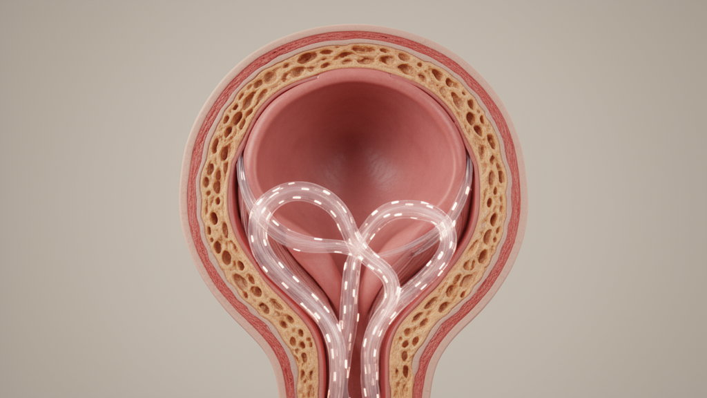
Researchers have addressed how histological findings within the intersphincteric region of the anal canal relate to contemporary surgical concepts, emphasizing that proposed connections remain hypotheses. Writing in response to clinical interpretations by Clemente and Khaw, the research team addressed proposed anatomical frameworks, including the described Garg fascia, outer-sphincteric space, and specialized RIFIL fistula pathways. The authors emphasized the essential distinction between structures directly demonstrated in normal cadaveric specimens and pathological fistula trajectories inferred from radiological imaging and operative observations.
The underlying anatomical investigation demonstrated that the human intersphincteric region possesses a structurally heterogeneous architecture. Microscopic examinations identified dense and loose longitudinal muscle components arranged alongside branching loose fibers traversing interbundle gaps within the external anal sphincter. In addition, histological preparations demonstrated a layered organization of the levator ani muscle.
Despite this observed complexity, the investigators clarified that their histological study did not specifically define the proposed Garg fascia. Furthermore, the investigation did not directly demonstrate an outer-sphincteric space or RIFIL pathways within normal cadaveric tissue. Consequently, the authors caution that connecting these specific surgical concepts to normal microscopic anatomy represents an anatomically plausible and testable hypothesis rather than an established histological reality.
To resolve these questions, the authors propose integrating high-resolution histology with magnetic resonance imaging and operative findings in patients with complex anal fistulas. Such future investigations may clarify these proposed pathways. This may support more precise, sphincter-preserving treatment.
Subject of Research: Histological anatomy of the anal canal intersphincteric region and proposed clinical fistula pathways
Article Title: Histological anatomy and clinical fistula pathways: reply to Clemente and Khaw
Article References: Muro, S., Nakajima, Y., Nimura, A., & Akita, K. (2026). Histological anatomy and clinical fistula pathways: reply to Clemente and Khaw. International Journal of Colorectal Disease, 41(1), Article 189. https://doi.org/10.1007/s00384-026-05272-x
Image Credits: AI Generated
DOI: 10.1007/s00384-026-05272-x
Keywords: anal canal, intersphincteric region, histological anatomy, anal fistula, fistula pathways, Garg fascia, outer-sphincteric space, RIFIL fistulas, external anal sphincter, levator ani, longitudinal muscle, sphincter-preserving surgery, cadaveric specimens, magnetic resonance imaging, colorectal disease, surgical anatomy
News Source: Ophelia Keating. (November 19, 2026). Anal Intersphincteric Region Is Structurally Heterogeneous While Fistula Pathways Remain Hypotheses. Scienmag.