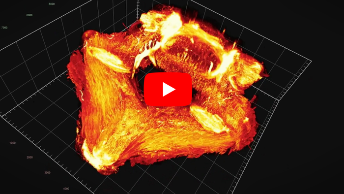
Back pain affects many people at some point in their lives, and a common cause is damage to the squishy discs or flexible, rubbery tissues of the spine. However, observing this damage at an early stage is difficult with current imaging methods. Now, researchers reporting in ACS Nano can see microscopic soft tissue destruction in animal spines by targeting denatured collagen with fluorescent molecules. Watch a video of the spinal imaging technique here.
Credit: Video credit belongs to the American Chemical Society.
Back pain affects many people at some point in their lives, and a common cause is damage to the squishy discs or flexible, rubbery tissues of the spine. However, observing this damage at an early stage is difficult with current imaging methods. Now, researchers reporting in ACS Nano can see microscopic soft tissue destruction in animal spines by targeting denatured collagen with fluorescent molecules. Watch a video of the spinal imaging technique here.
Anywhere along the spine, from the neck to tail bone, can become uncomfortable when its soft and protective tissues, including the cartilage and jelly-like intervertebral discs, become damaged and lose their structure. Daily wear-and-tear, as well as some disorders, such as facet joint osteoarthritis or ankylosing spondylitis, can degrade and unfurl the collagen proteins that give these tissues their bounce and flexibility. Detecting compromised collagen early could help patients get relief before the pain becomes severe, but this is very difficult to do with existing medical technologies, such as X-rays and magnetic resonance imaging (MRI). Previously, Yang Li and colleagues developed a collagen hybridizing peptide (CHP) probe that specifically binds unfurled collagen molecules, which happens when they deteriorate and lose their ability to cushion vertebrae. So, Li, Kuibo Zhang, Hong Shan and colleagues wanted to test if CHP labeled with fluorescent tags could be used as an imaging method to identify collagen destruction in the body.
To make the peptide probe more stable in the body, the researchers modified CHP by substituting a hydroxyl group with fluorine and then attaching a fluorescent dye to it. When healthy mice and rats were injected with the fluorescent dye-labeled CHP and imaged with near-infrared fluorescence (NIRF), the team could confirm that the fluorescing molecules accumulated on the soft tissues between the vertebrae. Then the researchers removed a portion of the animals’ spines and imaged them with light sheet fluorescence microscopy. This technique produced precise 3D maps, which revealed denatured collagen. Because CHP is known to specifically target damaged collagen, the team says their imaging experiments show that even healthy animals can have a modest degree of deteriorated collagen around load-bearing joints, especially in the lower back. In additional experiments, both the NIRF images and 3D maps generated with the new method detected collagen deterioration in animal models of spinal injury before structural changes were visible in tissues on MRI scans. Finally, the researchers applied dye-labeled CHP as a stain to intervertebral disc slides from people that had undergone spinal surgeries. The fluorescence intensity of the stain rose substantially as the level of disc degeneration increased. Based on these results, the researchers say that their molecular-level technique could be developed in clinical studies for earlier diagnosis and targeted therapeutic treatments for patients with back pain.
The authors acknowledge funding from the National Key R&D Program of China; the National Natural Science Foundation of China; the 2017 & 2018 High-level Health Team of Zhuhai; and the COVID-19 Emerging Prevention Products, Research Special Fund of Zhuhai City.
The paper’s abstract is available here.
The American Chemical Society (ACS) is a nonprofit organization chartered by the U.S. Congress. ACS’ mission is to advance the broader chemistry enterprise and its practitioners for the benefit of Earth and all its people. The Society is a global leader in promoting excellence in science education and providing access to chemistry-related information and research through its multiple research solutions, peer-reviewed journals, scientific conferences, eBooks and weekly news periodical Chemical & Engineering News. ACS journals are among the most cited, most trusted and most read within the scientific literature; however, ACS itself does not conduct chemical research. As a leader in scientific information solutions, its CAS division partners with global innovators to accelerate breakthroughs by curating, connecting and analyzing the world’s scientific knowledge. ACS’ main offices are in Washington, D.C., and Columbus, Ohio.
To automatically receive news releases from the American Chemical Society, contact [email protected].
Follow us: Twitter | Facebook | LinkedIn | Instagram
Journal
ACS Nano
DOI
10.1021/acsnano.1c07112
Article Title
“Molecular Imaging of Collagen Destruction of the Spine”
Article Publication Date
5-Nov-2021


