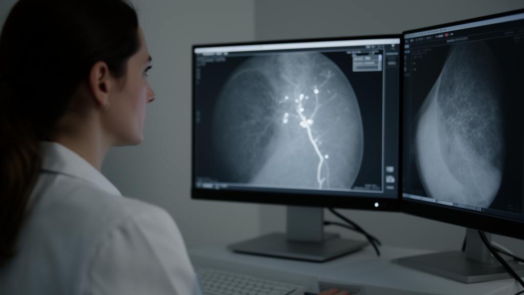
Every year, tens of millions of mammograms are performed worldwide, and many of them contain a quiet clue about a woman’s heart health that goes unreported. Breast arterial calcifications, the calcium deposits that build up in the walls of the arteries running through breast tissue, are a frequent incidental finding on routine mammograms. They correlate with coronary artery calcification and vascular events in women, and cardiovascular disease remains the leading cause of death in women worldwide, surpassing all cancers combined. Yet despite this well-documented association, BAC scoring is rarely implemented in clinical practice. Surveys indicate that while 60 to 81 percent of radiologists recognize BAC as a cardiovascular risk marker, only 16 to 62 percent document it consistently. With breast imaging workloads rising, researchers have been searching for scalable ways to extract this additional value from images that already exist, without any extra imaging or radiation exposure.
A new study published in European Radiology Breast reports that an artificial intelligence system can detect these calcifications automatically in a large real-world screening population, with very high specificity but only moderate sensitivity. The retrospective single-center study, conducted at the University Hospital of Zurich and its affiliated outpatient clinics in Switzerland, included 4692 women who underwent 6061 digital full-field mammographic examinations comprising 24,519 images between 2011 and 2017. The median age at first mammography was 57 years, and women with BAC were significantly older than those without, at 68 versus 56 years. The researchers set out to evaluate the diagnostic performance of an AI-based BAC detection system and to assess, for the first time, how technical and image-quality parameters influence automated detection in routine mammography.
The AI tool, an experimental module included in the b-box plus software package by b-rayZ, evaluated all mammographic images and classified each finding as vascular or non-vascular. The deep-learning model was trained on standard 2D full-field digital mammography and 2D synthetic tomosynthesis images from public and private datasets spanning multiple clinical sites in various countries and different manufacturers, with experienced breast radiologists annotating all training images. The data was split into a training set of 70 percent, a validation set of 20 percent, and a test set of 10 percent. The model was applied to standard craniocaudal and mediolateral oblique views, assigning a binary BAC score to each image and each patient.
Because manually reviewing every image would have been impractical, a representative subset of 375 patients, comprising 1993 images, was randomly selected for expert re-evaluation. A board-certified radiologist undergoing subspecialty training in breast imaging performed a focused BAC assessment according to predefined criteria and established the reference standard for this subset. In this manually reviewed subset, the human reader identified BAC in 349 images, or 17.5 percent, while the AI identified BAC in 144 images, or 7.2 percent. The model achieved an overall accuracy of 0.88 and an excellent specificity of 0.99, but sensitivity was limited at 0.35. The positive predictive value was high at 0.85, and the negative predictive value reached 0.88. At the patient level, 95 of the 375 women, or 25.3 percent, had BAC on at least one mammogram.
When the AI and the radiologist disagreed, the pattern of errors was revealing. Of the 247 discordant images, only 21 cases, or 8.5 percent, were false positives in which the AI indicated BAC but the radiologist did not. These discrepancies appeared most frequently associated with postoperative changes such as implants, surgical clips, scars, or architectural distortion, although in 3 of the 21 cases the AI assessment suggested the presence of BAC that may have been overlooked by the radiologist. False negatives were far more common, comprising 226 cases in which the radiologist identified BAC but the AI did not. These were most commonly associated with very subtle or low-intensity calcifications, often involving only a single vessel wall or discontinuous calcified foci with a tubular configuration, accounting for 42.9 percent of missed cases. Dense breast tissue potentially obscuring vascular structures was another frequent contributing factor, at 33.6 percent.
Across the full cohort, AI detected BAC in 652 of 4692 women, or 13.9 percent, in 800 of 6061 examinations, or 13.2 percent, and in 1644 of 24,519 images, or 6.7 percent. The authors caution that these analyses address factors influencing AI-based detectability of BAC rather than true differences in BAC prevalence. In multivariable mixed-effects logistic regression models accounting for within-patient clustering, compression force emerged as the most consistent technical factor associated with AI-detected BAC across projections, with significant associations in both craniocaudal and mediolateral oblique views on both sides of the body. Radiation dose showed a significant association only in left mediolateral oblique projections. Post-surgical status was strongly associated with BAC detection in the right craniocaudal projection, with an odds ratio of 5.06, while no significant associations were observed in the remaining projections.
Strikingly, most acquisition-related image-quality parameters showed no significant independent association with detection. Glandular profile, pectoralis visibility, inframammary fold depiction, nipple-to-pectoralis line correctness, breast elevation, and absence of skin folds were not independently associated with BAC detection. Breast density categories were not significantly associated with BAC presence in any projection or laterality, although dense breast tissue frequently contributed to false-negative classifications in qualitative review. The authors suggest that the higher compression and lower dose seen in BAC-positive craniocaudal images likely reflect easier compression in predominantly fatty, postmenopausal breasts, which allows adequate image quality at lower doses and may facilitate AI-based detection. Overall, the algorithm’s robustness to moderate technical variability suggests that even suboptimal images can still contribute meaningfully to automated BAC detection.
The study has important limitations that shape how the results should be read. Its retrospective single-center design may limit generalizability, and the reference standard was based on visual expert assessment available for only a subset of images, meaning subtle calcifications may have been missed or misclassified by the reader. No correlation with coronary CT, adverse cardiovascular events, or cardiovascular outcomes was performed, so no prognostic conclusions can be drawn from this study. The analyses of image-quality parameters were observational, and observed associations should not be interpreted as causal effects on BAC presence. No adjustments for age or imaging vendor were applied in the statistical models, and the study focused exclusively on standard two-view digital mammography, leaving performance in tomosynthesis and other modalities unknown. The authors also note that the algorithm is not suitable as a standalone screening tool, and negative results should be interpreted with caution, given that a substantial proportion of BAC cases are not detected.
Despite these constraints, the findings carry real clinical weight. The authors argue that the algorithm’s prioritization of specificity over sensitivity may be advantageous in opportunistic cardiovascular screening settings where minimizing false positives is critical, because highly specific algorithms are particularly valuable for reliably identifying patients with definite BAC. Very recently, AI-based BAC quantification has demonstrated incremental prognostic value beyond established cardiovascular risk scores and identifies BAC as a strong, independent predictor of adverse cardiovascular events. Future research, the authors suggest, should focus on automated BAC quantification to establish an Agatston-like scoring system and define clinically meaningful thresholds for initiating cardiovascular evaluation, given that higher calcification burden appears to be associated with increased risk. Given the high volume of screening examinations, AI-based opportunistic BAC detection could serve as a scalable and clinically practical entry point for targeted cardiovascular risk assessment and preventive care, allowing scalable identification of women with definite vascular calcifications who may benefit from targeted cardiovascular evaluation.
The study also fills a knowledge gap that has long gone unaddressed. Prior work established quality criteria to optimize cancer detection in mammography, but as BAC is a secondary finding, the requirements for image-quality parameters potentially affecting the visibility of calcified vessels were not fully understood. Positioning errors have been shown to be the single most important factor influencing final image quality, and inadequate positioning has been shown to reduce the sensitivity of mammographic assessment by up to 21 percent compared with properly positioned examinations. Technical image quality has also been shown to influence the detection of calcifications, with reduced microcalcification detection reported at higher image noise and lower radiation doses. That acquisition-related parameters were not directly associated with BAC detection by the AI in this study suggests these factors primarily influence detectability rather than the underlying presence of BAC. As the recently updated BI-RADS lexicon explicitly acknowledges the reported association between BAC and increased cardiovascular risk, tools that reliably flag these deposits from images already being acquired may bring a long-recognized but underused risk marker one step closer to routine clinical use.
Subject of Research: Diagnostic performance of an AI system for detecting breast arterial calcifications on mammography and the influence of image-quality parameters in a real-world screening population.
Article Title: AI-enabled detection of breast arterial calcifications in mammography: diagnostic performance and impact of image quality in a large real-world screening population
Article References: Saenger, J., Weber, A., Nikolova, E., Dudle, A., Happe, J., Schrading, S., Frauenfelder, T., & Boss, A. (2026). AI-enabled detection of breast arterial calcifications in mammography: diagnostic performance and impact of image quality in a large real-world screening population. European Radiology Breast, 1(1), Article 3. https://doi.org/10.1007/s44502-026-00003-y
Image Credits: AI Generated
DOI: 10.1007/s44502-026-00003-y
Keywords: breast arterial calcifications, mammography, artificial intelligence, deep learning, cardiovascular risk, image quality, screening, sensitivity, specificity, opportunistic screening, breast density, digital mammography
News Source: Ophelia Keating. (October 28, 2026). AI Spots Breast Artery Calcifications on Routine Mammograms with High Specificity but Modest Sensitivity. Scienmag.