In a groundbreaking pilot study, researchers have developed an automatic image co-registration technique that synergizes Carotid Angiography and Intravascular Optical Coherence Tomography (OCT) employing sophisticated machine learning methodologies. This innovative approach marks a significant advancement in the medical imaging field, focusing on enhancing the precision of cardiovascular diagnostics and treatment planning. The study propounds that integrating various imaging modalities can provide a comprehensive view of vascular health, thereby supporting clinicians in making more informed decisions.
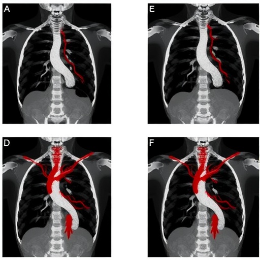
Carotid Angiography, a widely used imaging technology, offers detailed visualizations of blood vessels in the head and neck. Coupled with the high-resolution imaging capability of OCT, physicians can gain crucial insights into the structural and functional aspects of arterial walls. However, aligning these different imaging techniques has traditionally posed a considerable challenge. The advent of machine learning algorithms provides a way to overcome the limitations of manual co-registration, enhancing both accuracy and efficiency of the combined imaging approach.
The research presented by Xu et al. delves deeper into this revolutionary method, elaborating on the algorithms implemented to automate the co-registration process. By leveraging deep learning techniques, the researchers trained their models on a substantial dataset, enabling the algorithm to learn the complexities of different imaging modalities. The results reveal an impressive ability of the machine learning models to accurately align the images, demonstrating higher fidelity than conventional methods.
In clinical settings, the ability to seamlessly integrate these images could lead to better diagnosis and monitoring of cardiovascular diseases. Carotid artery disease, for instance, is a significant contributor to stroke, making accurate assessment critical. The newly developed automated image registration can potentially facilitate longitudinal assessments of disease progression or treatment efficacy, enriching the patient care pathway.
The study also highlights the methodological rigor employed in validating the effectiveness of the machine learning approach. The researchers utilized quantitative performance metrics to evaluate the accuracy and reliability of the co-registered images. This rigorous validation process not only underscores the robustness of their findings but also holds promise for broader applications in medical imaging beyond just carotid studies.
While the findings exhibit considerable potential, the authors also acknowledge the limitations of the pilot study. For instance, the sample size was relatively small, meaning that further research with larger cohorts is necessary to confirm these initial results. Additionally, the complexity of biological systems may pose additional challenges in diverse patient populations, particularly with varying anatomical features that may require fine-tuning of the model.
Despite these challenges, the implications of this research are far-reaching. The automatic co-registration technique can significantly reduce the time clinicians spend on image preparation, allowing them to focus on interpretation and decision-making regarding patient care. Moreover, this innovation aligns with a broader trend in medicine — the increasing reliance on artificial intelligence and machine learning to enhance clinical practices.
Moreover, the automatic nature of this process could lower the barrier to entry for smaller medical facilities that may lack access to expensive imaging software capable of performing manual alignments. By democratizing accessibility to advanced cross-sectional imaging analyses, the study holds the promise of improving health outcomes on a wider scale, particularly in underserved regions.
As the study underscores the mounting evidence in favor of adopting machine learning solutions, it also fuels the ongoing discussion around the regulatory and ethical frameworks necessary for integrating AI in healthcare. Due to the profound implications for patient care, incorporating AI in medical systems must be handled with utmost caution, ensuring that the technology is not only effective but also safe for patients.
Looking forward, the researchers express a desire to continue refining their algorithms and expanding the scope of their studies. They envision future applications wherein the co-registration technique could be adapted to other vascular regions or even different organ systems altogether, allowing for further exploration of the intricate relationships between structure and function in human health.
In conclusion, Xu et al.’s pioneering work encapsulates the essence of modern healthcare innovation — maximizing the potential of technology to enhance diagnostic practices. As we embrace this era of machine intelligence in medicine, studies like these pave the path for improved integration of diagnostic imaging, thereby transforming how clinicians approach complex cardiovascular conditions.
Harnessing the power of machine learning for automated image registration not only enhances current clinical practices but also opens avenues for future research aimed at unveiling new truths about human health and disease. As researchers continue to innovate, we anticipate a future where such technological advancements become standard practice, revolutionizing patient care.
As the worlds of technology and medicine converge, we remain optimistic about what lies ahead, as each new study brings us one step closer to realizing the full potential of artificial intelligence in enhancing human health.
Subject of Research: Automatic image co-registration using machine learning techniques in conjunction with Carotid Angiography and Intravascular Optical Coherence Tomography.
Article Title: Automatic Image Co-registration of Carotid Angiography and Intravascular Optical Coherence Tomography Based on Machine Learning Method: A Pilot Feasibility Study.
Article References:
Xu, H., Li, JN., Xu, Y. et al. Automatic Image Co-registration of Carotid Angiography and Intravascular Optical Coherence Tomography Based on Machine Learning Method: A Pilot Feasibility Study.
Ann Biomed Eng (2025). https://doi.org/10.1007/s10439-025-03872-2
Image Credits: AI Generated
DOI: https://doi.org/10.1007/s10439-025-03872-2
Keywords: Machine Learning, Image Co-registration, Carotid Angiography, Intravascular Optical Coherence Tomography, Cardiovascular Imaging.
Tags: AI image co-registrationalgorithms for image alignmentautomated image analysis techniquescardiovascular diagnostics innovationCarotid Angiography advancementsdeep learning for medical applicationsenhancing diagnostic accuracyIntravascular Optical Coherence Tomographymachine learning in medical imagingmulti-modal imaging integrationprecision medicine in cardiologyvascular health assessment technologies
