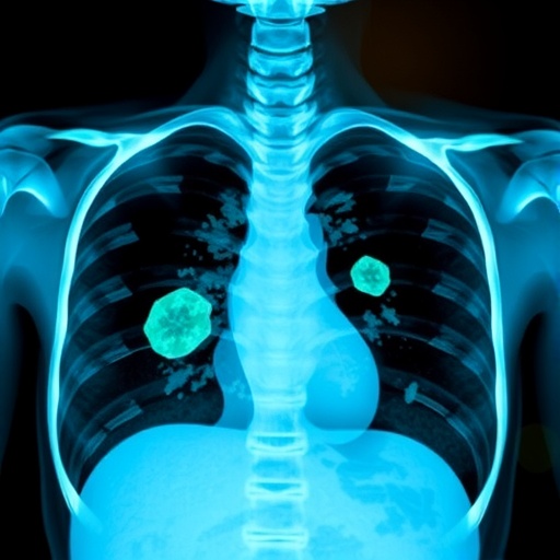
In a groundbreaking advancement at the intersection of artificial intelligence and medical imaging, researchers at the University of Southampton have engineered an AI-powered diagnostic tool capable of detecting subtle foreign objects lodged within patients’ airways with unprecedented accuracy. These objects, often invisible even to expert radiologists upon routine CT scans, present a formidable clinical challenge due to their radiolucent nature, which causes them to blend indistinguishably with surrounding anatomical structures.
Radiolucent foreign body aspiration (FBA) typically occurs when small objects, such as food particles or organic materials like plant matter and crustacean shells, inadvertently enter the respiratory tract. Their elusive imaging signature often leads to delayed or missed diagnoses, escalating the risk of severe respiratory distress or chronic complications. Despite the proficiency of seasoned radiologists, the intrinsic difficulty in visualizing these foreign bodies results in a diagnostic sensitivity that remains suboptimal. It is within this clinical conundrum that the deep learning model developed by the Southampton team demonstrates transformative potential.
Harnessing the power of deep neural networks, the researchers devised a composite AI model integrating a state-of-the-art airway segmentation framework known as MedpSeg, coupled with a convolutional neural network (CNN) meticulously trained on chest CT imaging data. This model was exposed to over 400 cases spanning multiple independent cohorts, ensuring robustness and diverse representation. The approach capitalizes on the model’s ability to parse subtle textural and morphological variances imperceptible to the human eye, discerning anomalies suggestive of foreign material entrapment within bronchial passages.
The research employed rigorous validation protocols, pitting the AI’s diagnostic acumen against the interpretations of three expert radiologists, each boasting over a decade of clinical experience. The evaluation comprised 70 CT scans, wherein 14 were confirmed cases of radiolucent FBA validated through bronchoscopy — the current gold standard for diagnosis. Intriguingly, while radiologists exhibited impeccable precision by avoiding false positives, they detected only a mere 36% of actual FBA cases, underscoring the intrinsic challenge of human-led visual assessment under these conditions.
By contrast, the AI system achieved a detection sensitivity of 71%, effectively reducing the margin of undiagnosed cases. Although the model’s precision registered at 77%, indicating some false positive signals, its overall diagnostic balance as measured by the F1 score reached an impressive 74%, significantly surpassing the radiologists’ 53%. This metric synthesizes both recall and precision, highlighting the AI’s superior capability to flag potential FBA cases for further clinical scrutiny without overwhelming practitioners with spurious alerts.
Underpinning this breakthrough is an advanced image analysis pipeline that meticulously maps the intricate bronchial architecture, enabling the system to localize and evaluate suspicious regions within the airways. The unique MedpSeg technique facilitates high-precision airway segmentation, serving as a foundational step that enhances the downstream neural network’s focus and classification accuracy. By training the AI to recognize characteristic patterns associated with radiolucent foreign bodies, the researchers circumvent traditional imaging limitations that have long impeded prompt and accurate diagnosis.
The implications of this research reach far beyond incremental diagnostic improvements. Timely identification and intervention in FBA cases are critical, as undetected foreign bodies can provoke inflammatory responses, airway obstruction, recurrent infections, and potentially irreversible lung damage. An AI-assisted diagnostic adjunct promises not only to enhance clinical workflow efficacy but also to markedly improve patient outcomes by minimizing diagnostic uncertainty and expediting treatment decisions.
Dr. Yihua Wang, the study’s lead author, emphasized that their AI tool is designed to operate in synergy with radiologists, augmenting rather than replacing human expertise. “Our model acts as a vigilant second observer, offering additional assurance in complex cases where traditional imaging interpretations are prone to oversight,” Wang elucidated. This collaborative approach fosters a paradigm shift toward augmented intelligence in medical practice, where computational precision complements clinical judgment.
Looking ahead, the research team is poised to expand validation efforts via multicenter trials involving larger, more demographically varied populations to further refine the model’s accuracy and generalizability. Reducing potential algorithmic biases inherent in training datasets remains a priority to ensure equitable performance across diverse patient cohorts. The ultimate goal is to translate this technology into widespread clinical adoption, transforming how respiratory foreign body aspirations are detected and managed globally.
This pioneering work has been meticulously documented in the recent publication titled Automated Detection of Radiolucent Foreign Body Aspiration on Chest CT Using Deep Learning, featured in the journal npj Digital Medicine. The study is underpinned by collaborative efforts between the University of Southampton and clinical partners in Wuhan, China, and benefits from support by the UK Medical Research Council and the China Scholarship Council, symbolizing a potent international alliance in advancing medical AI.
In conclusion, this AI-driven innovation heralds a new frontier in medical imaging diagnostics, where machine learning enhances the detection capabilities for complex, otherwise elusive pathologies. By addressing critical gaps in radiological diagnosis with superior sensitivity and balanced precision, the technology holds the promise to save lives and reduce healthcare burdens, setting a precedent for future AI applications in medical science.
Subject of Research:
People
Article Title:
Automated detection of radiolucent foreign body aspiration on chest CT using deep learning
News Publication Date:
10-Nov-2025
Web References:
https://www.nature.com/articles/s41746-025-02097-w
References:
Automated Detection of Radiolucent Foreign Body Aspiration on Chest CT Using Deep Learning, npj Digital Medicine, DOI: 10.1038/s41746-025-02097-w
Image Credits:
University of Southampton
Keywords:
Artificial intelligence, Medical diagnosis, Medical imaging, Imaging, Medical tests, Clinical imaging, Health and medicine, Radiology
Tags: advanced convolutional neural networks in radiologyAI in medical imagingAI vs human radiologistsairway segmentation in imagingchest scan analysis with AIclinical challenges in detecting foreign bodiesdeep learning in healthcarediagnostic accuracy in respiratory medicineforeign body aspiration detectionmachine learning for medical diagnosticsradiolucent foreign objects in CT scansSouthampton University AI research


