In a groundbreaking advancement poised to revolutionize interventional nephrology, researchers Wang, Calle, Yan, and colleagues have introduced a pioneering technique that employs a convolutional neural network (CNN)-based optical coherence tomography (OCT) endoscope to guide percutaneous nephrostomy procedures. This novel approach melds cutting-edge artificial intelligence with high-resolution imaging, offering unparalleled precision and safety in the placement of nephrostomy catheters, a vital intervention for patients suffering from urinary tract obstructions and related renal complications.
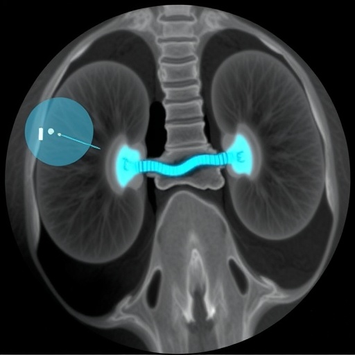
Percutaneous nephrostomy involves the insertion of a catheter into the renal pelvis to drain urine directly from the kidney in cases where normal passage through the ureter is blocked. Traditionally, this procedure relies heavily on fluoroscopy or ultrasound imaging guidance, methods that, despite their widespread use, pose limitations in terms of spatial resolution and tissue differentiation. The integration of OCT technology, known for its micron-level resolution and capability to capture cross-sectional images of biological tissues, marks a dramatic improvement in the visualization of renal structures during catheter placement.
At the heart of this innovation is the utilization of a convolutional neural network—a form of deep learning algorithm adept at pattern recognition in complex imaging data. The CNN was meticulously trained to interpret OCT images in real-time, discriminating between various tissue types and anatomical landmarks within the renal system. This intricate AI-enabled image analysis enables the endoscope not only to provide a detailed visual pathway but also to offer predictive insights that enhance the clinician’s ability to maneuver the catheter safely and efficiently.
The convolutional neural network’s prowess stems from its layered architecture, allowing it to extract hierarchical features from raw OCT data. Through successive convolutional layers, the network identifies nuances in tissue texture and composition, such as differentiating healthy renal parenchyma from fibrotic or inflamed regions. Incorporating training datasets from a diverse patient cohort ensured the CNN’s robustness across variable anatomical presentations and pathological conditions.
Optical coherence tomography itself works on the principle of low-coherence interferometry, projecting near-infrared light into tissue and measuring the time delay and intensity of backscattered light. The adaptation of this technology into a miniaturized endoscope provides volumetric imaging capabilities within the narrow confines of the nephrostomy tract, overcoming the spatial limitations inherent to traditional imaging. This compact OCT probe, integrated with the CNN, can deliver near-instantaneous 3D maps of internal renal architecture, which were previously unattainable during nephrostomy.
Clinically, the implications are profound. By providing a detailed and real-time anatomical roadmap, this integrated system minimizes the risk of inadvertent injury to surrounding vasculature or adjacent organs, significantly reducing complication rates. Moreover, the enhanced visualization shortens procedure times, diminishes radiation exposure, and potentially increases the success rate of first-attempt catheter placements. These factors cumulatively contribute to improved patient outcomes and decreased healthcare costs.
The interdisciplinary nature of this development involved collaboration between biomedical engineers, computer scientists, and nephrologists, exemplifying a model for future technological innovation in medicine. The extensive validation phase included in vitro tissue models, animal studies, and initial human trials to confirm the safety, accuracy, and reproducibility of the OCT-CNN guided percutaneous nephrostomy. Results indicated superior performance metrics when compared against conventional imaging guidance methods, heralding a new standard for procedural precision.
Moreover, the authors anticipate that this technology could be extended beyond nephrostomy to other minimally invasive surgical interventions where tissue differentiation and navigation within delicate anatomical spaces are paramount. The adaptability of CNN-driven OCT endoscopy holds promise for applications in vascular surgery, gastroenterology, and oncology, where precise imaging is critical for targeted therapy.
Focus on real-time data processing was essential for the system’s clinical relevance. Latency in imaging feedback was minimized through optimized computational frameworks, allowing the convolutional neural network to analyze OCT images almost instantaneously. This responsiveness ensures that surgeons receive continuous guidance, making dynamic adjustments during the procedure possible, a feature not feasible with delayed or static imaging.
The study further explored the integration of user-friendly interfaces that translate the CNN-OCT data into intuitive visualizations for clinicians. Augmented reality overlays within the endoscopic view help direct catheter navigation while simplifying complex anatomical relationships. Such user-centered design not only enhances surgical precision but also reduces cognitive load during high-stress interventions.
Despite these advancements, the researchers acknowledge challenges ahead, including the need for broader clinical trials to confirm efficacy across varied patient populations and diverse clinical settings. Additionally, ongoing development focusing on miniaturization and cost reduction of OCT hardware will be essential to facilitate widespread adoption in healthcare systems worldwide.
Finally, the data security and ethical considerations surrounding AI in medical devices were addressed, highlighting the importance of transparent algorithmic design and patient data anonymization. Robust regulatory pathways must be established to ensure safety and build trust within the medical community regarding AI-assisted procedures.
In summary, Wang, Calle, Yan, and their team have delivered a transformative hybrid solution to percutaneous nephrostomy guidance, ushering in a new era where convolutional neural networks empower optical coherence tomography endoscopy. Their work exemplifies the future of precision medicine—leveraging artificial intelligence and advanced imaging to enhance clinical outcomes, minimize risks, and redefine standards in minimally invasive procedures.
Subject of Research:
Percutaneous nephrostomy guidance using convolutional neural network-integrated optical coherence tomography endoscopy.
Article Title:
Percutaneous nephrostomy guidance by a convolutional-neural-network-based optical coherence tomography endoscope.
Article References:
Wang, C., Calle, P., Yan, F. et al. Percutaneous nephrostomy guidance by a convolutional-neural-network-based optical coherence tomography endoscope. Commun Eng (2026). https://doi.org/10.1038/s44172-026-00613-8
Image Credits: AI Generated
Tags: advanced imaging for kidney interventionsAI and OCT integration in urologyAI-enhanced urinary tract obstruction treatmentAI-guided OCT endoscopeconvolutional neural network in nephrostomydeep learning for medical imaginghigh-resolution imaging in interventional nephrologyoptical coherence tomography for renal procedurespercutaneous nephrostomy catheter placementprecision nephrostomy guidancereal-time OCT image interpretationsafety improvements in nephrostomy techniques


