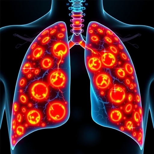
In a groundbreaking study that could redefine the landscape of oncology, researchers have unveiled an advanced AI-powered spatial cell phenomics platform designed to vastly improve risk stratification in non-small cell lung cancer (NSCLC). This novel approach harnesses the power of artificial intelligence to analyze the complex spatial organization and phenotypic diversity of cells within tumor microenvironments, providing unprecedented insights into tumor biology and patient prognosis. Given the global burden of NSCLC, which accounts for the majority of lung cancer cases and high mortality rates worldwide, innovations that enable precise risk assessment and personalized treatment strategies are of critical importance.
The study, spearheaded by Schallenberg, Dernbach, Ruane, and colleagues, presents a sophisticated integration of high-dimensional cell imaging and AI algorithms that collectively establish a comprehensive framework for spatial phenomics. Unlike traditional methods reliant on bulk tissue analysis or limited biomarker panels, this platform preserves spatial context at single-cell resolution, enabling clinicians and researchers to interpret how different cell populations interact and contribute to tumor progression. This shifts the paradigm from mere molecular profiling to a multidimensional understanding of tumor ecology, which is particularly vital in the heterogeneous landscape of NSCLC.
Central to this technology is the application of multiplexed imaging techniques that generate rich datasets capturing the expression of numerous markers within intact tumor sections. These detailed images serve as input for deep learning models trained to accurately identify and classify diverse cell phenotypes, including tumor cells, immune infiltrates, stromal components, and vascular elements. By delineating cellular neighborhoods and their compositional dynamics, the AI can detect subtleties in tumor architecture that correlate strongly with clinical outcomes. This level of resolution and contextual mapping surpasses conventional histopathological evaluation, opening avenues for more nuanced risk stratification.
The AI framework integrates multiple layers of data including cell morphology, protein expression profiles, and spatial distribution patterns to build predictive models capable of categorizing patients based on risk categories. By correlating these models with longitudinal clinical data sets, the researchers demonstrated that their approach significantly enhances the accuracy of prognosis prediction compared to existing staging systems. Importantly, this improvement was observed across diverse NSCLC cohorts, underscoring the robustness and generalizability of the platform.
One of the pivotal insights from this work is the identification of spatial biomarkers that represent patterns of cellular interactions predictive of aggressive disease phenotypes. For instance, certain configurations involving immune cell exclusion or localized immunosuppressive niches within the tumor microenvironment were linked with poorer survival outcomes. Traditional assessment methods often fail to capture such intricate spatial relationships, highlighting the advantage of AI-driven spatial phenomics in revealing tumor-immune crosstalk critical for therapeutic decision-making. This detailed characterization lays the groundwork for tailored immunotherapy regimens that consider not only the presence but also the spatial context of immune populations.
Beyond prognostic utility, the AI platform has potential applications in guiding therapeutic strategies by allowing the monitoring of spatial phenotypic changes in response to treatment. Given NSCLC’s heterogeneous response to chemotherapy, targeted therapies, and immunomodulators, real-time spatial phenomic profiling could offer dynamic biomarkers to predict and track treatment efficacy, resistance mechanisms, and disease recurrence. Such feedback mechanisms are invaluable for adaptive treatment protocols aiming to optimize patient outcomes while minimizing toxicity.
Technically, the study involved harnessing convolutional neural networks (CNNs) optimized for image segmentation and cell classification, trained on an extensive annotated dataset generated from multiplex immunofluorescence assays. The researchers employed rigorous cross-validation techniques and external cohort testing to validate the reproducibility and accuracy of their models. Furthermore, advanced clustering algorithms were utilized to reveal cellular communities and tissue structures correlating with clinical endpoints. The computational pipeline demonstrated scalability and potential integration with routine pathology workflows, emphasizing translational feasibility.
In addition to improving clinical decision-making, the findings provide fundamental biological insights into NSCLC pathogenesis. By mapping spatial heterogeneity and cellular phenotypes with high precision, the study uncovers previously unappreciated tumor microenvironmental niches that may harbor therapeutic vulnerabilities. This could catalyze future research focused on disrupting specific cell-cell interactions or modifying the tumor architecture to improve immunosurveillance and treatment responsiveness.
The implications of this research extend beyond NSCLC, heralding a new era in cancer diagnostics where AI-powered spatial phenomics becomes a cornerstone technique. The multi-parameter, high-resolution data combined with powerful machine learning analytics exemplifies a transformative methodology applicable to various tumor types characterized by complex microenvironments. As spatial omics technologies continue to evolve and become more widely accessible, integration of such AI frameworks promises to accelerate biomarker discovery, enhance patient stratification, and ultimately guide precision oncology approaches globally.
Importantly, the study acknowledges challenges related to data standardization, computational resources, and clinical implementation barriers. The authors advocate for collaborative efforts to develop harmonized protocols for sample preparation, imaging, and data analysis, which will be essential for multi-center validation and regulatory approval. Ethical considerations around AI transparency and interpretability in clinical contexts also require attention to ensure trust and adoption by pathologists and oncologists. Despite these hurdles, the authors remain optimistic that continued advancements and investments in AI-driven phenomics will revolutionize cancer diagnostics and personalized medicine.
This innovative research is poised to ignite a paradigm shift in how cancers are studied and treated, moving away from one-dimensional molecular markers toward holistic, spatially resolved characterizations of tumor ecosystems. The dynamic interplay of tumor cells with their microenvironment, captured at the single-cell level and powered by AI, reveals a novel dimension of cancer biology imperative for improving prognostic precision and therapeutic targeting. With lung cancer remaining a formidable global health challenge, this AI-powered spatial cell phenomics framework promises to enhance clinical practice and outcomes for millions of patients worldwide.
As the oncology community embraces the integration of artificial intelligence and spatial biology, this pioneering study provides a compelling proof-of-concept for the future of cancer risk stratification. By combining rich spatial context with sophisticated phenotype analysis, researchers and clinicians can now approach NSCLC with a new lens—one that not only identifies risk but also informs the design of personalized interventions. The convergence of machine learning and spatial imaging thus represents a vital frontier in the relentless pursuit of conquering cancer.
In sum, the work by Schallenberg et al. stands as a milestone in leveraging AI to decode the complexity of the tumor microenvironment and translate these insights into actionable clinical tools. This approach exemplifies the power of interdisciplinary innovation, merging computational science, pathology, and oncology to tackle one of the deadliest cancers. It provides a scalable and generalizable framework capable of transforming diagnostic pathology and patient care in NSCLC and potentially across the cancer spectrum.
The research also inspires further exploration into integrating spatial phenomics with other omics modalities, such as genomics and transcriptomics, to achieve even deeper multi-layered understanding of tumor biology. The future of oncology will likely be shaped by such comprehensive, AI-driven platforms, enabling truly personalized and adaptive therapeutic strategies, ultimately improving survival and quality of life for cancer patients globally.
This landmark study is a shining example of how AI and spatial biology synergize to unlock new dimensions of clinical and biological insights, paving the way for next-generation cancer diagnostics and therapeutics. As this technology matures and gains widespread clinical translation, it holds the promise to revolutionize cancer treatment paradigms and bring us closer to the goal of precision, predictive, and personalized oncology.
Subject of Research: Artificial intelligence-driven spatial cell phenomics applied to risk stratification in non-small cell lung cancer
Article Title: AI-powered spatial cell phenomics enhances risk stratification in non-small cell lung cancer
Article References:
Schallenberg, S., Dernbach, G., Ruane, S. et al. AI-powered spatial cell phenomics enhances risk stratification in non-small cell lung cancer. Nat Commun 16, 9701 (2025). https://doi.org/10.1038/s41467-025-65783-z
Image Credits: AI Generated
DOI: https://doi.org/10.1038/s41467-025-65783-z
Tags: AI spatial cell analysisartificial intelligence in oncologyheterogeneous tumor ecologyhigh-dimensional cell imaginglung cancer risk stratificationmultiplexed imaging techniquesnon-small cell lung cancer researchpatient prognosis in lung cancerpersonalized cancer treatment strategiesspatial phenomics platformtumor biology insightstumor microenvironment analysis


