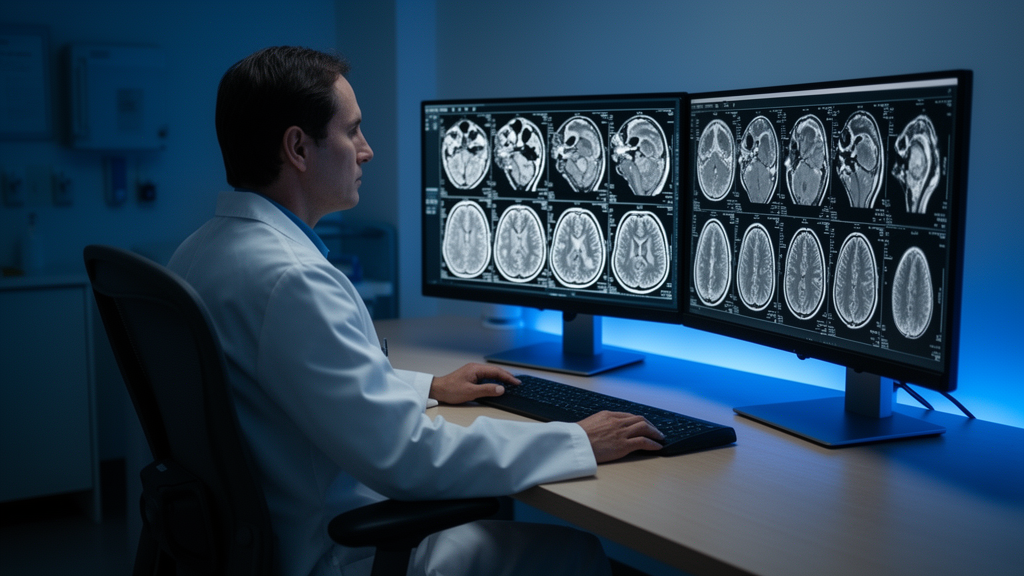
Spanish researchers evaluating artificial intelligence tools have found that deep-learning software can detect stroke damage in routine brain magnetic resonance imaging scans performed without contrast dye, though overall accuracy currently remains moderate. In a comparative evaluation of three distinct machine learning approaches, the self-configuring nnU-Net framework achieved the highest segmentation performance, while a lightweight model based on the YOLOv8 architecture followed closely behind. A conventional three-dimensional U-Net, long regarded as a benchmark in medical computer vision, lagged substantially behind both modern systems. Because the overall accuracy of the top models was moderate and lesion detection varied widely between scans, the investigators emphasized that these findings represent an exploratory initial step rather than an automated diagnostic system ready for hospital implementation.
Ischemic stroke constitutes a primary cause of mortality and long-term disability globally, making the rapid identification of damaged brain tissue critical for determining appropriate medical interventions. Brain scans utilizing fluid-attenuated inversion recovery, commonly known as FLAIR, can reveal tissue swelling and historical strokes without requiring patients to receive an intravenous contrast agent. Because FLAIR sequences are available on most standard clinical scanners, the technique presents an appealing basis for widespread or automated screening programs, particularly for individuals who cannot safely receive chemical contrast dye. Nevertheless, automated interpretation of FLAIR images remains technically challenging because stroke lesions can be small, faint, and irregular. Furthermore, acute lesions often closely resemble benign, age-related changes in the brain’s white matter or technical imaging artifacts, while hardware differences among scanners introduce further visual inconsistency.
To evaluate how fundamentally different computational architectures handle non-contrast brain scans under identical experimental conditions, the research team utilized the publicly available ISLES 2022 dataset. This medical imaging archive includes 250 stroke cases accompanied by expert-delineated lesion outlines. The authors trained all three machine learning models exclusively on FLAIR scans, applying identical data splits and uniform image preprocessing routines wherever feasible. Performance was evaluated across 25 independent test cases using the Dice similarity coefficient, a standard mathematical metric ranging up to 1.0, where a score of 1 represents a complete spatial overlap with the expert radiologist outlines.
The investigation examined three distinct engineering strategies: a manually configured 3D U-Net representing a traditional medical segmentation baseline, the self-configuring nnU-Net system that automatically tunes its internal settings to match dataset properties, and YOLOv8, a high-speed system originally designed for everyday object detection. The investigators adapted YOLOv8 to process volumetric brain scans slice by slice before algorithmically rebuilding the individual two-dimensional detections into a unified three-dimensional output. In testing, nnU-Net achieved an average Dice score of 0.483, outperforming the baseline 3D U-Net score of 0.294 by approximately 64 percent. Meanwhile, YOLOv8 delivered an average Dice score of 0.476, matching the leading self-configuring system across many test cases despite its simpler underlying architecture.
The comparative analysis also highlighted clear qualitative and structural differences among the outputs produced by each algorithm. The nnU-Net model consistently generated smoother and more coherent anatomical boundaries, successfully delineating irregularly shaped stroke lesions. In contrast, while YOLOv8 performed nearly as well in aggregate, it tended to miss stroke lesions that were unusually small, diffuse, or faint against surrounding brain tissue. In addition, all three machine learning models displayed substantial performance variation from scan to scan. Notably, even the top-performing nnU-Net architecture failed to identify any stroke lesion whatsoever in several test cases.
The study authors concluded that nnU-Net represents the superior option when spatial accuracy and boundary consistency are the primary priorities and sufficient computing resources are available. Conversely, YOLOv8 offers a viable, practical alternative when processing simplicity and screening speed take precedence, such as in initial diagnostic triage. The traditional 3D U-Net remains a valuable historical benchmark for comparative evaluations. However, the researchers cautioned that several methodological constraints limit immediate clinical translation. The experimental validation was conducted on a relatively small testing sample of 25 cases, and computational constraints permitted only a single training run, or fold, of the demanding nnU-Net model.
Additional limitations noted by the team include the absence of external validation on magnetic resonance scans acquired from other hospital facilities. Furthermore, while YOLOv8 possesses an inherent structural speed advantage stemming from its lightweight two-dimensional design, the authors did not formally measure or quantify processing times during the study. The experiments also relied strictly on FLAIR sequences, deliberately excluding other magnetic resonance imaging sequences contained within the ISLES 2022 dataset that could provide complementary anatomical detail. Given these factors, the researchers characterized their work as an exploratory benchmark and recommended future evaluations involving larger and more heterogeneous datasets.
Looking ahead, the authors suggested that future research should examine multi-sequence magnetic resonance protocols and explore hybrid computational systems that integrate the spatial precision of three-dimensional architectures with the operational speed of two-dimensional models. If perfected, dependable contrast-free artificial intelligence screening could assist radiologists in detecting subtle vascular damage, enhance diagnostic consistency across clinical shifts, and minimize reliance on contrast agents in vulnerable patient populations. The research was led by corresponding author Patricia García-Berlanga, Juan Zapata, and Juan Martínez-Alajarin from the Universidad Politécnica de Cartagena, alongside Pablo Hernandez-Cerdan from Vasomaly, with research funding provided by Spain’s Ministry of Science, Innovation and Universities.
Subject of Research: Artificial intelligence detection of stroke lesions in non-contrast brain magnetic resonance imaging
Article Title: Detection of vascular pathologies in non-contrast magnetic resonance images for early screening
Article References: Detection of vascular pathologies in non-contrast magnetic resonance images for early screening. Original publication (Bibliographic details unavailable.)
Image Credits: AI Generated
DOI: Not provided
Keywords: ischemic stroke, magnetic resonance imaging, FLAIR sequence, nnU-Net, YOLOv8, 3D U-Net, lesion segmentation, ISLES 2022 dataset, Dice score, non-contrast MRI, deep learning, stroke screening, Universidad Politécnica de Cartagena, Patricia García-Berlanga, medical image analysis, brain tissue damage, Journal of Intelligent Systems in Current Computer Engineering, Bentham Science Publishers
News Source: Cassandra Pierce. (November 12, 2026). AI Detects Stroke Damage in Non-Contrast Brain MRI With Moderate Accuracy. Scienmag.