In a groundbreaking advancement combining artificial intelligence with medical imaging, researchers at Osaka Metropolitan University have developed an AI-based diagnostic tool capable of detecting esophageal achalasia using plain chest X-rays. This achievement promises a less invasive, more accessible, and potentially earlier diagnosis for a disorder that historically demands complex procedures. The innovation addresses a critical clinical challenge, marking a significant leap forward in non-invasive diagnostic accuracy.
Esophageal achalasia is a rare but serious disorder characterized by the impaired relaxation of the lower esophageal sphincter and disrupted peristalsis, leading to difficulties in food passage from the esophagus into the stomach. Patients typically report symptoms such as dysphagia, regurgitation of undigested food, chest pain, and significant weight loss. Traditional diagnosis relies heavily on invasive methods including high-resolution manometry and endoscopy, which though effective, are resource-intensive and uncomfortable for patients.
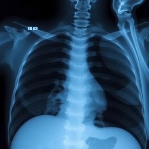
Chest radiography has long been used as a preliminary imaging tool, primarily to exclude other thoracic conditions. However, diagnosing achalasia purely through plain X-rays has been elusive due to the subtlety and variability of radiographic features like esophageal dilation, twisting, or fluid retention. The standard practice often necessitates barium swallow studies to visualize esophageal motility, adding complexity and exposure to contrast agents. This new AI-driven approach directly challenges these limitations by leveraging subtle imaging biomarkers invisible to the human eye.
The team at Osaka Metropolitan University, comprising Dr. Tadashi Ochiai, Dr. Akinari Sawada, and Associate Professor Daiju Ueda, harnessed deep learning algorithms trained with a comprehensive dataset consisting of 207 chest X-rays from 144 achalasia patients and 240 chest X-rays from matched controls without the disease. This dataset meticulously captured a spectrum of disease presentations, allowing the AI to learn nuanced features beyond conventional radiographic interpretation.
Validation of the model employed a distinct test set of 17 achalasia cases and 64 non-achalasia controls, rigorously ensuring the AI’s diagnostic capability was robust across different patient samples. The model achieved remarkable accuracy metrics including an Area Under the Curve (AUC) of 0.964, sensitivity of 94.1%, and specificity of 89.1%. These values translate into a diagnostic tool that can detect most true cases while minimizing false positives, outperforming physicians’ diagnostic performance under similar conditions.
A remarkable component of this research was the use of heatmap visualizations to interpret the AI model’s decision process. These overlays highlight the esophageal region prominently, particularly focusing on dilation and structural abnormalities. This transparency is crucial not only for clinician trust but also for elucidating the pathological hallmarks that might be overlooked or underestimated in manual assessment. Through this explainable AI framework, the traditionally opaque “black box” of machine learning gains medical credibility.
The clinical implications of this research extend beyond diagnostic accuracy. Esophageal achalasia is notoriously difficult to diagnose promptly; studies indicate an average delay of approximately 6.5 years from symptom onset to confirmed diagnosis. This delay exacerbates esophageal dilation and tortuosity, complicating treatment and reducing patient outcomes. Early detection using a widely available, minimally invasive tool like chest X-ray analysis could revolutionize patient care paradigms by facilitating timely intervention.
In Japan, where chest X-rays are routinely performed as part of general health screenings, integrating this AI diagnostic system could enable mass screening for achalasia. This represents a paradigm shift in early diagnostics for esophageal motor disorders, emphasizing accessibility and prevention. The AI’s non-reliance on specialized equipment or invasive protocols means broader population-level surveillance with minimal patient burden.
From a technological standpoint, the successful application of AI to plain radiography underscores the untapped potential of existing imaging modalities. By unlocking richer diagnostic data from simple tools, healthcare systems can leverage AI innovations to optimize workflows, reduce costs, and improve outcomes with lower barriers to implementation. The Osaka Metropolitan University team’s methodology reflects a convergence of clinical insight with state-of-the-art machine learning practices.
Furthermore, this study contributes to the growing body of evidence that AI can surpass traditional diagnostic limitations. The incorporation of subtle radiographic patterns into computational models hints at a future where many ‘invisible’ pathological markers may become readily diagnosable. This could transform not only gastrointestinal medicine but also broader diagnostic radiology and clinical decision-making.
While the current model’s performance is impressive, ongoing research and larger multicenter trials will be vital to validating these results across diverse populations and healthcare contexts. Future integration with clinical data, symptomatology, and other imaging modalities may further enhance diagnostic accuracy and risk stratification capabilities.
The researchers emphasize that this AI system is a supplementary tool designed to aid, not replace, physicians. Its most valuable role may be as an initial screening modality guiding further confirmatory testing. By blending AI’s precision with expert clinical judgement, the early detection and management of achalasia—and potentially other esophageal motility disorders—could enter a new era.
In sum, the innovative AI application developed by Osaka Metropolitan University showcases how intelligent algorithms can repurpose standard diagnostic images, enabling earlier, less invasive detection of esophageal achalasia. This breakthrough shines as a beacon for future AI-driven advancements within medical diagnostics, demonstrating extraordinary promise to reshape clinical practice patterns, improve patient outcomes, and reduce healthcare burdens associated with late diagnoses.
—
Subject of Research: People
Article Title: Artificial Intelligence-Based Detection of Achalasia on Plain Chest Radiography
References: Clinical Gastroenterology and Hepatology
Image Credits: Osaka Metropolitan University
Keywords: Health and medicine, Diagnostic imaging, Diagnostic accuracy, Medical diagnosis
Tags: AI diagnostic tools for achalasiaArtificial Intelligence in Medicinechest X-ray advancementsclinical challenges in achalasiaearly diagnosis of achalasiaesophageal achalasia detectionimproving diagnostic accuracy with AImedical imaging innovationsnon-invasive imaging techniquespatient-friendly achalasia diagnosisreducing discomfort in achalasia diagnosistraditional vs non-invasive diagnosis
