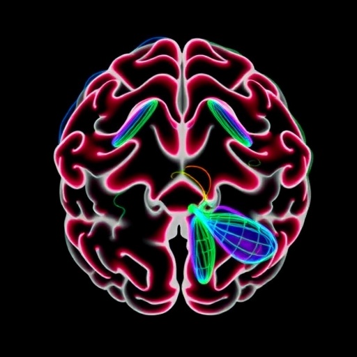
In a groundbreaking study published in Nature Communications, a multinational team of neuroscientists sheds new light on how aging constrains the brain’s spatial geometry, revealing profound implications for our understanding of cognitive decline and neural plasticity. This research pioneers a comprehensive approach combining advanced neuroimaging, cutting-edge mathematical modeling, and large-scale data analytics, opening fresh avenues for investigating how age-related physical changes in neural architecture relate directly to functional deterioration across the lifespan.
The human brain, a labyrinth of intricate networks and geometrical configurations, undergoes myriad transformations as we age. While previous research has largely emphasized molecular and cellular markers of aging, this new study shifts the paradigm by focusing on spatial geometry—the shape, curvature, and folding patterns of brain structures—and how these physical properties constrain information processing capabilities. Escalante and colleagues explore these age-related spatial constraints in unprecedented detail, drawing from a rich dataset comprising over a thousand individuals spanning from early adulthood to advanced age.
Central to their approach is the innovative use of topological data analysis and geometric morphometrics, methodologies often employed only in abstract mathematical fields or physical sciences, but rarely applied to neuroscience at this scale. By quantifying the brain’s surface geometry and volume distribution, the researchers reveal systematic alterations in spatial organization that do not merely reflect degradation but impose fundamental limits on brain functionality. This reframing of aging as a geometric phenomenon holds transformative potential for diagnostic and therapeutic strategies.
What emerges from the data is a clear pattern: the brain’s spatial geometry becomes increasingly constrained with age. Structures that in young brains exhibit high complexity and dynamic flexibility, such as the cerebral cortex’s gyrification patterns and white matter tracts’ connectivity, progressively lose their intricate foldings and optimal spatial configurations. This loss of geometric complexity disrupts efficient neural communication pathways, suggesting a physical basis for the cognitive deficits observed in aging populations.
Further analysis unveils that these geometric constraints affect not only gray matter regions responsible for executive functioning, memory, and sensory processing but also the distribution and integrity of white matter fibers that facilitate rapid signal transmission. Reduced curvature and altered spatial topology correlate with diminished connectivity strength and slower cognitive processing speeds, underscoring the multi-level impact of spatial degradation.
The study also challenges the traditional viewpoint that cortical thinning or volume loss alone accounts for cognitive decline. Even when controlling for these volumetric changes, geometric constraints remain a significant predictor of reduced cognitive performance, indicating that spatial organization is an independent and critical factor. This insight compels a rethink of neural aging, highlighting the brain’s three-dimensional structure as equally vital to its functional preservation.
Escalante et al. integrate these findings with longitudinal cognitive assessments, linking geometric alterations to specific behavioral outcomes such as impaired spatial navigation, reduced working memory capacity, and slowed executive control functions. This integrative perspective provides a more cohesive understanding of how physical and physiological aging converge to impair cognition, potentially guiding tailored interventions that aim to preserve spatial integrity.
One particularly exciting aspect of this work is the identification of early geometric biomarkers that precede significant cognitive symptoms. Changes in local curvature and spatial distribution were detected in individuals years before clinical diagnoses of mild cognitive impairment were established. This temporal predictive power introduces the possibility of earlier intervention strategies designed to maintain or restore optimal brain geometry.
Moreover, the researchers offer a theoretical model elucidating how age-related biochemical processes, such as altered extracellular matrix composition and cytoskeletal degradation, may mechanistically precipitate these geometric constraints. This model bridges the gap between microstructural cellular changes and macrostructural brain morphology, marrying biological aging with physical transformations at a level not previously appreciated.
The implications extend toward neuroplasticity research as well. If brain geometry imposes fundamental constraints, understanding how the aging brain reorganizes spatially could inform rehabilitation paradigms following injury or neurodegenerative disease. The study prompts inquiry into whether targeted activities or pharmaceuticals might mitigate or reverse detrimental geometric shifts, preserving neural circuit functionality.
Technologically, this research marks a milestone in brain imaging. Utilizing ultra-high-field MRI combined with novel computational algorithms, the team captures spatial data with remarkable resolution and accuracy. These methodological advances not only enhance reproducibility and precision but also set a new standard for future neuroimaging studies exploring structural-functional relationships.
Ethical and societal ramifications also arise. With improved diagnostic capacity based on spatial biomarkers, debates concerning screening, early detection, and personalized aging interventions will intensify. Balancing benefits with privacy, access, and psychological impacts remains a challenge for integrating such advanced knowledge into clinical practice.
In sum, this landmark investigation redefines brain aging as a fundamentally geometric phenomenon. By spotlighting spatial constraints as key drivers of cognitive decline, Escalante et al. encourage a holistic, multi-scale understanding of the aging brain that transcends volume loss to incorporate shape, connectivity, and topological integrity. Their work paves the way for innovative diagnostics, enhanced predictive capacity, and transformative therapies targeting the physical foundations of neural aging.
As the global population ages, unraveling these spatial mechanisms is paramount. The convergence of mathematics, biology, and technology embodied in this study offers hope for sustaining brain health and cognitive vitality well into late life, challenging assumptions and illuminating new horizons in brain science.
Subject of Research: Age-related alterations in the spatial geometry of the brain and their impact on cognitive function.
Article Title: Age-related constraints on the spatial geometry of the brain.
Article References:
Escalante, Y.Y., Adams, J.N., Yassa, M.A. et al. Age-related constraints on the spatial geometry of the brain. Nat Commun 16, 8613 (2025). https://doi.org/10.1038/s41467-025-63628-3
Image Credits: AI Generated
Tags: advanced methodologies in brain researchage-related physical changes in the brainaging impacts on cognitive declinebrain spatial geometry and agingfunctional deterioration across the lifespangeometric morphometrics in neuroscienceimplications of aging on brain architectureintricate networks of the human brainlarge-scale data analytics in neuroscienceneural plasticity and ageneuroimaging techniques in neurosciencetopological data analysis in brain studies


