Recent advancements in medical imaging technology have become critical in the battle against cancer, particularly in the early detection of recurrent cases. A groundbreaking study led by researchers including Baltacioglu, M.H., Soydal, C., and Araz, M., explores the enhanced capabilities of whole abdomen FDG PET/MRI scans in comparison to the standard whole body PET/CT for identifying peritoneal recurrence in ovarian cancer patients. This research is pivotal for clinicians and patients alike, as it sheds light on more accurate diagnostic methods, potentially leading to improved clinical outcomes.
Ovarian cancer is notoriously difficult to detect early, which significantly hampers treatment effectiveness. Many patients initially respond well to treatment but suffer recurrences due to metastatic spread to the peritoneum, making early detection of such recurrences crucial. Traditional imaging techniques, including PET/CT, have been the cornerstone of staging and follow-up care in patients with ovarian cancer. However, emerging methodologies like FDG PET/MRI are beginning to show promise in providing additional anatomical and functional information, which could significantly influence management strategies.
In the study conducted by Baltacioglu and colleagues, researchers aimed to compare the diagnostic accuracy of the whole abdomen FDG PET/MRI with standard whole body PET/CT scans specifically for assessing peritoneal recurrence. The incorporation of MRI not only allows for detailed imaging of soft tissues but, when coupled with functional PET data, gives insight into metabolic activities of tumors. This fusion of anatomical and metabolic imaging technologies is revolutionary, as it could enable healthcare professionals to visualize and characterize peritoneal lesions more effectively.
The study utilized a cohort of ovarian cancer patients who were previously treated and were under surveillance for possible recurrence. Participants underwent both imaging techniques, and the results were meticulously analyzed to ascertain which modality provided superior detection rates of peritoneal metastases. The findings indicated that whole abdomen FDG PET/MRI significantly outperformed standard PET/CT, highlighting the benefits of MRI’s high-resolution imaging capabilities in revealing small and subtle lesions that might otherwise be missed.
One of the critical advantages of FDG PET/MRI lies in its reduced radiation exposure compared to conventional imaging methods. This aspect is especially important for cancer patients who often require multiple imaging sessions throughout their treatment journey. The ability to achieve high diagnostic accuracy without subjecting patients to excessive radiation doses represents a major leap forward. This is particularly relevant considering the long-term effects of radiation exposure in cancer survivors, who may already face an elevated risk of developing secondary malignancies.
Furthermore, the metabolic information provided by FDG PET enhances the specificity of lesions detected through MRI. The study posited that the metabolic activity of peritoneal lesions could correlate strongly with the biological aggressiveness of the tumors. As such, the integration of PET with MRI not only improves the likelihood of detecting cancer recurrence but also aids in refining treatment planning and potentially prognostic assessments of patients.
The implications of these findings could be far-reaching. If adopted into standard clinical practice, the enhanced diagnostic capabilities of whole abdomen FDG PET/MRI could ensure earlier and more accurate intervention strategies, which are vital for improving survival outcomes in ovarian cancer patients. The potential to tailor treatment regimens based on precise imaging insights represents a significant advancement in personalized medicine.
In addition, the findings contribute to the growing body of evidence supporting the shift towards hybrid imaging technologies in oncology. As the field of cancer diagnosis continues evolving, it’s essential for clinicians and researchers to embrace these innovations that allow for enhanced patient care. The research team’s work is a testament to the ongoing commitment to advancing cancer imaging techniques, ultimately aiming to improve the quality of life for patients battling this formidable disease.
The successful application of whole abdomen FDG PET/MRI in this research setting opens doors for further studies to explore its efficacy across different cancer types, as well as its role in various stages of disease management. Future research initiatives should aim to investigate whether this imaging method can also be beneficial in detecting recurrences in other solid tumors, thus broadening its potential clinical implications.
Stakeholders in the healthcare system, including policymakers and insurance providers, should take note of the evidence surrounding the effectiveness and safety of whole abdomen FDG PET/MRI. Establishing guidelines for reimbursement and accessibility will be crucial to ensuring that this transformative imaging technology can reach all patients in need, making it a standard tool in the oncology imaging arsenal.
As the research continues to be scrutinized, the ultimate goal remains the same: to arm physicians with the best tools available for fighting cancer. The promising results presented in this study suggest a pivotal shift in how recurrences of ovarian cancer may be detected in the future, with an emphasis on accuracy and patient safety.
In summary, the exploration of whole abdomen FDG PET/MRI versus standard whole body PET/CT offers exciting new insights into the early detection of peritoneal recurrence in ovarian cancer. As research in this area progresses, the hope is that these innovations will lead to enhanced survival rates and improved quality of life for individuals affected by this devastating disease. Enhanced imaging techniques could very well be a game-changer in the ongoing battle against cancer, reaffirming the importance of research and development in the medical field.
Subject of Research: Detection of peritoneal recurrence of ovarian cancer using imaging techniques.
Article Title: Additive value of whole abdomen FDG PET/MRI to standard whole body PET/CT for detection of peritoneal recurrence of ovarian cancer.
Article References:
Baltacioglu, M.H., Soydal, C., Araz, M. et al. Additive value of whole abdomen FDG PET/MRI to standard whole body PET/CT for detection of peritoneal recurrence of ovarian cancer. J Ovarian Res (2026). https://doi.org/10.1186/s13048-025-01662-x
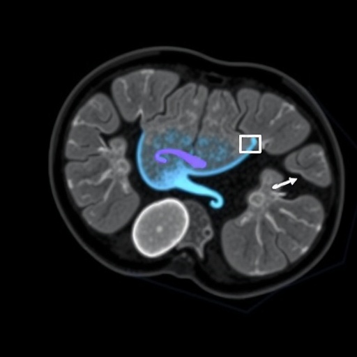
Image Credits: AI Generated
DOI:
Keywords: Ovarian cancer, PET/MRI, imaging techniques, peritoneal recurrence, diagnostic accuracy, personalized medicine, hybrid imaging.
Tags: cancer imaging advancementsclinical outcomes in cancer treatmentdiagnostic accuracy in oncologyearly ovarian cancer detectionFDG PET/MRI technologyimproved cancer management strategiesmetastatic spread in ovarian cancerMRI’s role in cancer diagnosisperitoneal recurrence detectionPET/CT imaging limitationsPET/MRI advantages in ovarian cancerwhole abdomen imaging techniques


