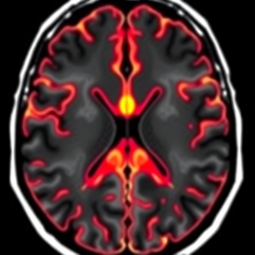
In a groundbreaking advancement that could reshape the way Parkinson’s disease is diagnosed and monitored, researchers have utilized sophisticated multiparametric quantitative magnetic resonance imaging (MRI) to reveal hitherto unseen microstructural changes in the putamen, a critical brain region affected by the disease. This study, recently published in npj Parkinsons Disease, harnesses cutting-edge neuroimaging techniques that go far beyond conventional MRI scans, offering profound insights into the subtle alterations in brain tissue composition and organization that occur in early to advanced stages of Parkinson’s disease. The implications of these findings promise not only to enhance diagnostic precision but also to pave the way for more individualized therapeutic strategies.
Parkinson’s disease, a progressive neurodegenerative disorder, primarily impacts the motor system, leading to tremors, rigidity, and bradykinesia. Traditionally, diagnosis has relied heavily on clinical symptoms and, when available, dopaminergic imaging such as dopamine transporter single-photon emission computed tomography (DaT-SPECT). However, these approaches offer limited resolution regarding the microstructural context of the underlying neuropathology. The innovative use of multiparametric quantitative MRI addresses this gap by enabling noninvasive, in vivo mapping of brain tissue properties at a microscopic scale and providing quantitative metrics that reflect pathological changes more directly.
Central to the research is the putamen, a subcortical structure in the basal ganglia, which plays a pivotal role in motor control and learning. In Parkinson’s disease, degeneration of dopaminergic neurons severely disrupts the functional circuitry of the basal ganglia, with the putamen being one of the earliest and most affected sites. By applying multiple quantitative MRI parameters—such as T1 and T2 relaxation times, magnetic susceptibility, and diffusion metrics—the team could dissect the complex microstructural environment of the putamen. These parameters essentially serve as biomarkers, each sensitive to different tissue characteristics, including iron deposition, myelin integrity, and cellular density.
.adsslot_Ai5WM6mOfZ{ width:728px !important; height:90px !important; }
@media (max-width:1199px) { .adsslot_Ai5WM6mOfZ{ width:468px !important; height:60px !important; } }
@media (max-width:767px) { .adsslot_Ai5WM6mOfZ{ width:320px !important; height:50px !important; } }
ADVERTISEMENT
One of the notable aspects of this multiparametric approach is its capacity to differentiate between various pathological substrates within the putamen, which was previously impossible with standard MRI. For example, iron accumulation in basal ganglia structures is a known hallmark of Parkinsonian pathology and can exacerbate oxidative stress leading to neuronal death. By quantifying magnetic susceptibility values, the study demonstrates increased iron deposits localized within the putamen of Parkinson’s patients compared to healthy controls. This provides a compelling objective measure to track disease progression correlated with iron-mediated neurodegeneration.
In addition to iron mapping, the research emphasizes changes in water molecule diffusion patterns within the putamen’s microenvironment, acquired through diffusion tensor imaging (DTI) and related modalities. These diffusion metrics indicate alterations in tissue architecture, such as axonal damage or demyelination, which alter the directionality and magnitude of water diffusion. The study reveals reduced fractional anisotropy and increased mean diffusivity, signifying microstructural disruption and a loss of organized neural pathways within affected regions. These disruptions are thought to underlie motor deficits seen in Parkinson’s patients, linking imaging findings with clinical symptomatology.
Another essential quantitative parameter explored is the longitudinal (T1) and transverse (T2) relaxation times. Variations in these values reflect changes in tissue composition and molecular environment. The study uncovers significant prolongation of T1 and T2 times in the putamen, which may indicate neuroinflammatory processes and gliosis—responses to neuronal injury that contribute to the pathophysiology of Parkinson’s disease. Such markers open new avenues for understanding the inflammatory dimension of the disease, which had been challenging to assess without invasive procedures or histological analysis.
This multiparametric strategy also benefits from advanced image processing and machine learning algorithms that integrate these multiple MRI-derived contrasts into comprehensive microstructural maps. These computational tools enhance the sensitivity and specificity of detecting pathological changes, allowing for single-subject-level diagnostics that could revolutionize clinical practice. The study team reports high accuracy in discriminating Parkinson’s disease patients from healthy individuals, suggesting immediate translational potential for personalized medicine.
The longitudinal nature of the research provides further insights into disease trajectory. By following patients over time, the researchers demonstrate that microstructural alterations in the putamen evolve predictably with disease progression, correlating with worsening motor scores and functional impairment. This temporal dimension could enable clinicians to monitor treatment efficacy more objectively and adjust interventions before irreversible neurological damage ensues.
Technically, the research pushes the boundaries of MRI hardware and sequence design. High-field magnets, optimized pulse sequences, and meticulous calibration procedures were employed to improve signal-to-noise ratio and minimize imaging artifacts. Such technical rigor is essential to achieve the reproducibility and reliability of multiparametric quantitative MRI required for clinical adoption. The study sets a new standard for future neuroimaging investigations into Parkinson’s disease and other neurodegenerative disorders.
Clinically, these findings have profound implications. Early detection of microstructural changes before overt clinical symptoms manifest could enable intervention at a stage when neuroprotective therapies are more likely to be effective. Moreover, identifying specific pathological components such as iron overload or neuroinflammation could guide tailored therapeutic strategies, including chelation therapy or anti-inflammatory agents, potentially altering disease course.
Looking ahead, the integration of multiparametric quantitative MRI with other biomarkers—genetic, biochemical, or electrophysiological—may provide a holistic framework for comprehensive Parkinson’s disease profiling. Such multidimensional precision medicine approaches will ultimately improve patient outcomes by enabling bespoke treatments based on individual pathophysiology rather than one-size-fits-all paradigms.
The study also acknowledges limitations and challenges inherent to implementing this approach widely. As sophisticated imaging protocols require high-end MRI scanners and expertise, disseminating this technology globally might face logistical hurdles. Furthermore, normative data across diverse populations need establishment to account for biological variability. Nevertheless, continuous technological advances and growing clinical demand suggest these challenges are surmountable.
In summary, the employment of multiparametric quantitative MRI to uncover microstructural putamen changes represents a transformative leap in Parkinson’s disease research. It redefines our ability to visualize and quantify intricate pathological processes noninvasively with remarkable detail. This technological milestone holds the promise of earlier diagnosis, refined disease monitoring, and targeted therapeutic development, ultimately improving quality of life for millions affected by Parkinson’s disease worldwide.
As neuroscience and imaging technology converge, studies like this exemplify the power of interdisciplinary collaboration to decode complex brain disorders. The insights gained enrich our fundamental understanding of Parkinson’s disease and equip clinicians with novel tools to combat its devastating effects. The future of neurodegenerative disease management looks more hopeful than ever, driven by innovation at the intersection of physics, biology, and medicine.
With ongoing research, the scope of multiparametric quantitative MRI is poised to expand, encompassing not only Parkinson’s disease but other disorders characterized by microstructural brain changes, such as Alzheimer’s disease, multiple sclerosis, and Huntington’s disease. The paradigm shift toward comprehensive brain tissue characterization is ushering in a new era of diagnostic precision and personalized care.
Ultimately, this pioneering work underscores the transformative potential of advanced imaging in unraveling the complex pathophysiological tapestry of Parkinson’s disease. It invites the medical community to reimagine diagnostic criteria and therapeutic algorithms through the lens of microstructural neuroimaging biomarkers, heralding a future where neurological diseases are detected earlier, understood better, and treated more effectively than ever before.
Subject of Research: Microstructural changes in the putamen in Parkinson’s disease revealed by multiparametric quantitative MRI.
Article Title: Multiparametric quantitative MRI uncovers putamen microstructural changes in Parkinson’s disease.
Article References:
Drori, E., Cohen, L., Arkadir, D. et al. Multiparametric quantitative MRI uncovers putamen microstructural changes in Parkinson’s disease. npj Parkinsons Dis. 11, 197 (2025). https://doi.org/10.1038/s41531-025-01020-0
Image Credits: AI Generated
Tags: Advanced MRI techniquesbrain tissue composition analysisDaT-SPECT limitationsdiagnostic precision in Parkinson’searly-stage Parkinson’s detectionindividualized therapeutic strategiesmicrostructural changes in putamenmultiparametric quantitative MRIneurodegenerative disorder researchneuroimaging advancementsnoninvasive brain mappingParkinson’s disease diagnosis


