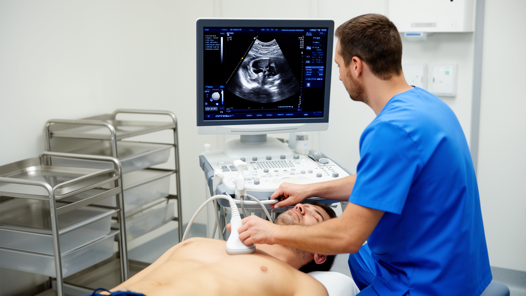
Cardiovascular medicine has historically focused on the left ventricle, often treating the right ventricle and tricuspid valve as hemodynamically subordinate structures of secondary physiological importance. Decades of early experimental studies and clinical observations reinforced this perception, showing that disabling the right ventricle in animal models caused no significant rise in venous pressure, while surgical procedures bypassing the chamber yielded favorable early results in congenital heart disease. However, contemporary cardiovascular science has fundamentally revised this perspective, establishing right ventricular dysfunction and tricuspid regurgitation as powerful independent predictors of morbidity and mortality across diverse cardiac conditions. Severe tricuspid regurgitation is no longer viewed merely as an innocent bystander or passive consequence of downstream disease, but rather as an active driver of adverse structural remodeling. In a comprehensive review published in Cardiovascular Ultrasound, Martijn G. H. Vrijkorte and colleagues synthesize modern diagnostic evidence to demonstrate that conventional imaging metrics systematically fail to capture the true functional status of the right ventricle, underscoring the urgent necessity of multiparametric, advanced echocardiographic evaluation.
Understanding the diagnostic challenges of the right ventricle requires careful examination of its unique embryological origins, anatomical architecture, and physiological properties. Arising from precursor cells located in the anterior heart field of the pharyngeal mesoderm, the right ventricle is morphologically and functionally distinct from the left ventricle, which originates from the first heart field. Structurally, the right ventricle forms a thin-walled crescentic chamber measuring between three and five millimeters in myocardial thickness that partially wraps around the thicker, spherical left ventricle. Anatomically divided into an inlet encompassing the tricuspid valve apparatus, a heavily trabeculated apical myocardium, and a smooth-walled infundibulum or outflow tract, its mechanics depend heavily on myocardial fiber orientation. Subendocardial longitudinal fibers running from the tricuspid annulus to the apex generate approximately eighty percent of systolic shortening, while outer circumferential fibers continuous with the left ventricle account for twenty-five percent of the wall and drive radial contraction. Contraction of the infundibulum contributes another fifteen percent of total stroke volume, typically occurring twenty to fifty milliseconds after apical contraction to generate a smooth, peristaltic ejection into the low-resistance pulmonary circuit.
Despite possessing only thirty percent of the myocardial mass of the left ventricle, the right ventricle matches its stroke volume while expending approximately half the energy per unit of generated force. This exceptional mechanical efficiency stems from its compliant geometry and substantial systolic ventricular interdependence, whereby left ventricular contraction augments up to thirty percent of right ventricular stroke output through shared fibers and septal motion. Conversely, diastolic ventricular interdependence means that severe right ventricular dilation within the non-compliant pericardial space directly restricts left ventricular filling, compromising overall cardiac output. These unique architectural features make the right ventricle exceptionally sensitive to changes in preload and afterload. In chronic volume overload states such as tricuspid regurgitation, the right ventricle initially deploys compensatory eccentric hypertrophy, Frank-Starling heterometric adaptation, and afterload-induced homeometric contractile augmentation via the Anrep effect. Over time, persistent hemodynamic stress exhausts these compensatory reserves, triggering a self-reinforcing vicious cycle of annular dilation, progressive dysfunction, systemic congestion, and organ failure, which Santas and colleagues characterized as a four-grade continuum that demonstrates a graded association with mortality in heart failure patients.
Conventional two-dimensional transthoracic echocardiography remains the primary first-line diagnostic modality due to widespread availability and bedside convenience, yet it is severely constrained by geometric assumptions and regional motion bias. Guidelines from the Tricuspid Valve Academic Research Consortium define severity thresholds for right ventricular dysfunction across standard metrics, classifying tricuspid annular plane systolic excursion as mild at fourteen to seventeen millimeters, moderate at ten to thirteen millimeters, and severe below ten millimeters. Tissue Doppler systolic velocity is graded as mild at nine to eleven centimeters per second, moderate at six to eight, and severe below six centimeters per second, while fractional area change is categorized as mild between thirty-four and thirty-seven percent, moderate between thirty and thirty-three percent, and severe below thirty percent. However, standard apical four-chamber views cannot visualize the right ventricular outflow tract, which accounts for twenty-five percent of total chamber volume, and frequently foreshorten the true ventricular apex. Consequently, these planar measurements provide an incomplete evaluation that frequently mischaracterizes global right ventricular contractile performance.
The clinical consequences of relying on isolated two-dimensional indices become particularly evident when comparing conventional parameters directly against volumetric reference standards. Tolvaj and colleagues evaluated conventional parameters across a large multicenter cohort and documented alarming misclassification rates reaching up to forty-nine percent for fractional area change when evaluated against three-dimensional ejection fraction. Strikingly, patients who were reclassified by three-dimensional ejection fraction below forty-five percent as having right ventricular dysfunction despite displaying completely normal conventional metrics experienced an all-cause mortality risk elevated more than fourfold. Among traditional measures, free wall longitudinal strain exhibited the strongest agreement with three-dimensional ejection fraction, achieving an area under the receiver operating characteristic curve of 0.877, though clinical outcomes remained poorest when patients presented with impairment across two or more parameters. Furthermore, isolated reductions in tricuspid annular plane systolic excursion frequently mask preserved radial function, whereas simultaneous reductions in both planar excursion and fractional area change identify advanced global failure and convey substantially worse long-term survival prospects.
Three-dimensional echocardiography eliminates geometric assumptions by providing direct volumetric quantification, achieving functional and volumetric accuracy comparable to cardiac magnetic resonance imaging while offering greater speed and clinical accessibility. Specialized software systems utilizing knowledge-based reconstruction can assemble reliable three-dimensional ventricular models from conventional two-dimensional imaging views and anatomical landmarks, reaching feasibility rates between eighty-five and ninety percent under optimized imaging protocols. Cardiac magnetic resonance remains the volumetric reference standard with superior tissue characterization, yet high costs, limited availability, claustrophobia, advanced renal dysfunction, cardiac devices, and an inability to calculate effective ejection fraction constrain its routine utility. Muraru and colleagues defined standardized three-dimensional reference ranges, categorizing ejection fraction as normal above forty-five percent, mildly reduced between forty and forty-five percent, moderately reduced between thirty and forty percent, and severely reduced below thirty percent. Multi-ethnic evaluations from the World Alliance of Societies of Echocardiography further revealed crucial biological differences, demonstrating that women maintain higher ejection fractions and strain values than men, while older individuals and Black subjects exhibit physiological shifts toward greater radial rather than longitudinal contraction.
Assessing diastolic performance and intrinsic myocardial contractility requires looking beyond conventional Doppler-derived filling indices such as the tricuspid inflow E/A ratio, E/e prime, and hepatic venous flow patterns, which remain highly preload-dependent and confounded by arrhythmias. Right atrial strain analysis of reservoir and conduit function has surfaced as a sensitive early indicator of diastolic failure, especially in atrial secondary regurgitation where the loss of coordinated atrial contraction compromises filling dynamics. To address the load dependence of systolic indices, non-invasive right ventricular myocardial work calculates pressure-strain relationships to assess myocardial performance, with global work efficiency and wasted work demonstrating stronger correlations with hemodynamic deterioration than traditional measures. In secondary regurgitation, Fabian and colleagues demonstrated that volume-adjusted global myocardial work indices independently predict mortality, while artificial intelligence models developed by Szijártó and colleagues reconstruct afterload-adjusted pressure curves directly from peak echocardiographic pressures. Additionally, continuous-wave Doppler evaluation of regurgitant jet acceleration provides right ventricular dP/dt measurements, where values exceeding 400 millimeters of mercury per second indicate preserved contractility and indexed dP/dt over maximal pressure outmatches unadjusted metrics across worsening functional classes.
Right ventricular to pulmonary arterial coupling describes the efficiency with which the right ventricle transfers energy to the pulmonary circulation, representing a critical determinant of right-sided circulatory integrity. The gold standard of coupling assessment requires invasive catheterization to calculate the ratio of end-systolic elastance to effective arterial elastance, with values below 1.0 defining pathological uncoupling that portends clinical decompensation. Echocardiographic surrogates, such as the ratio of tricuspid annular plane systolic excursion to pulmonary artery systolic pressure, offer non-invasive prognostic insights, yet regurgitant jets frequently underestimate pressure in severe regurgitation. To overcome these diagnostic hurdles, novel integrated formulas provide enhanced discrimination. A parameter integrating free wall longitudinal strain, three-dimensional end-diastolic volume, and invasive pulmonary artery systolic pressure achieved superior prognostic precision over traditional metrics, demonstrating a C-index of 0.68 versus 0.57 for one-year mortality after transcatheter interventions. Similarly, Ancona and colleagues identified a free wall strain to systolic pressure cutoff of 0.26 percent per millimeter of mercury that separates survivors from high-risk individuals, while the pulmonary artery pulsatility index identifies heightened risk when falling below four, and severe decompensation below two.
The clinical progression of tricuspid regurgitation is heavily shaped by underlying etiology, broadly divided by consensus into atrial and ventricular secondary phenotypes that manifest divergent remodeling patterns and prognostic profiles. Atrial secondary regurgitation arises primarily from right atrial enlargement and annular dilation in the setting of atrial fibrillation or heart failure with preserved ejection fraction, typically preserving longitudinal contractility and maintaining a favorable conical ventricular geometry. Conversely, ventricular secondary regurgitation stems from leaflet tethering, pulmonary hypertension, and primary myocardial disease, creating spherical midventricular remodeling that elevates wall stress, induces interventricular septal flattening, and produces the classic D-shaped left ventricle. Quantitative assessment by Tomaselli and colleagues revealed that an effective regurgitant orifice area exceeding 0.47 square centimeters doubles the combined risk of death or heart failure hospitalization, while three-dimensional imaging unmasks occult dysfunction in ostensibly low-risk patients. Contractile patterns provide vital insight into disease reversibility: compensated phases maintain ejection fraction through elevated circumferential shortening despite depressed longitudinal strain, whereas decompensated states marked by simultaneous longitudinal and circumferential failure carry one-year mortality rates approaching fifty percent, accompanied by elevated CA125 levels that outperform natriuretic peptides in severe volume overload.
The advent of transcatheter tricuspid valve interventions has fundamentally altered clinical management while introducing complex periprocedural imaging considerations that demand meticulous preprocedural evaluation. Acute elimination of the low-impedance regurgitant leak abruptly lowers right ventricular preload while increasing effective afterload, frequently inducing transient reductions in planar excursion, fractional area change, and free wall strain that reflect loading normalization or annular device splinting rather than true myocardial injury. Successful repair promotes beneficial reverse remodeling characterized by an initial biphasic reduction in end-diastolic volume followed by delayed end-systolic contraction and effective ejection fraction recovery. Nevertheless, clinical implementation faces persistent barriers, as suboptimal acoustic windows yield analyzable three-dimensional datasets in only fifty percent of studies, and fewer than twenty percent of clinicians routinely utilize advanced modalities despite equipment availability in ninety percent of laboratories. Closing these translational gaps through standardized protocols, artificial intelligence, and evidence-based thresholds remains essential to optimizing intervention timing and improving outcomes.
Subject of Research: Advanced echocardiographic assessment of right ventricular structure and function in tricuspid regurgitation
Article Title: Beyond the left ventricle: modern echocardiographic assessment of right ventricular function in tricuspid regurgitation
Article References: Vrijkorte, M. G. H., Wunderlich, N. C., Duncan, A., Rana, B. S., Timmers, L., van der Pol, R., & Swaans, M. J. (2025). Beyond the left ventricle: modern echocardiographic assessment of right ventricular function in tricuspid regurgitation. Cardiovascular Ultrasound, 23(1), Article 24. https://doi.org/10.1186/s12947-025-00360-4
Image Credits: AI Generated
DOI: 10.1186/s12947-025-00360-4
Keywords: echocardiography, right ventricle, tricuspid regurgitation, three-dimensional imaging, myocardial strain, cardiovascular imaging, transcatheter intervention, ventricular-arterial coupling, Beyond, left, ventricle, modern
News Source: Ophelia Keating. (November 12, 2026). Advanced Echocardiography Unlocks Right Ventricular Function in Severe Tricuspid Regurgitation. Scienmag.