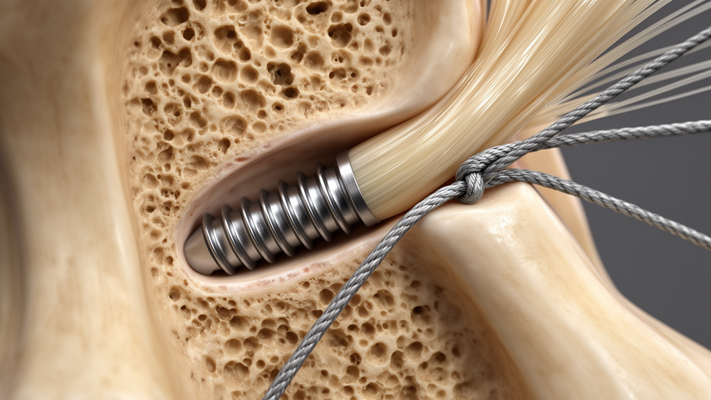
In an experimental investigation of anterior cruciate ligament reconstruction mechanics, supplementing standard tibial interference screws with high-strength sutures tied directly over an autologous cortical bone bridge produced substantial structural stability, matching the performance of costly commercial anchor devices while sharply curbing early mechanical failures under repetitive stress. Tibial fixation has long represented the primary structural vulnerability during the postoperative recovery phase following reconstructive knee surgery, particularly when surgeons utilize soft-tissue tendon grafts that lack an osseous plug. To address this persistent challenge without adding expensive implants or complex surgical hardware, an international research team evaluated whether tying graft-guiding sutures across a simple bridge carved into the anterior tibial cortex could reinforce conventional screw placement. Across 61 reconstructed porcine knee models, the combined hybrid construct sustained cyclic loading conditions and failure forces far superior to standalone techniques, establishing that basic anatomical bone bridges can deliver dependable biomechanical reinforcement.
Epidemiological data cited by the researchers underscores the widespread clinical impact of anterior cruciate ligament injuries, which occur at an estimated global incidence of approximately 70 cases per 100,000 person-years and concentrate heavily within athletic cohorts, with female athletes facing an elevated vulnerability. When active individuals undergo surgical reconstruction, hamstring autografts and comparable soft-tissue tendon configurations are frequently chosen, yet their biological incorporation into bone requires extended healing periods during which the mechanical security of the tibial tunnel serves as the sole barrier against joint laxity. While bioabsorbable interference screws designed to compress graft tissue against tunnel walls remain the clinical standard, single-point intratunnel fixation frequently encounters premature slippage when exposed to the accelerated physical therapy regimens favored in modern rehabilitation protocols. The senior clinical author had employed cortical suture tying over a bone bridge as routine clinical practice for fifteen years, prompting the formal multi-arm laboratory trial to systematically quantify its mechanical attributes alongside existing commercial alternatives.
The experimental design comprised a tightly controlled six-arm comparative biomechanical investigation utilizing a bioequivalent animal model consisting of fresh bovine digital extensor tendons mounted within porcine tibia specimens, a tissue pairing established in orthopedic literature for replicating human knee structural properties. A total of 61 test specimens were randomized across six distinct fixation configurations, with nine to eleven specimens allocated to each testing arm. Tendon grafts were carefully prepared by folding the extracted bovine tissue onto itself to generate bifascicular constructs with an average diameter of 8.5 millimeters, secured at both extremities using high-strength FiberLoop number two traction sutures woven in a quadruple braid configuration. Standardized tibial tunnels were drilled from the anteromedial tibial surface to the anatomical insertion of the anterior cruciate ligament on the anterior tibial spine at a fixed 55-degree angle using a 9-millimeter drill bit, faithfully reproducing the anatomical trajectory and dimensions typical of standard human surgical intervention.
To isolate the precise biomechanical contributions of each fixation strategy, the researchers compared two single-system models against four hybrid fixation approaches. Group one evaluated standalone cortical fixation, created by performing two converging 2-millimeter drill holes spaced one centimeter apart and one centimeter distal to the tibial tunnel aperture, passing the traction sutures through the resulting cortical bone bridge with a reverse needle and tying them securely. Group two examined standard isolated intratunnel fixation using a conical bioabsorbable BioComposite interference screw measuring 9 by 23 millimeters matching the drilled tunnel diameter. Group three combined the interference screw with suture tying across the drilled cortical bone bridge, while groups four, five, and six supplemented the interference screw with commercial secondary devices: a metallic cortical screw equipped with a toothed washer functioning as a post, a 4.75-millimeter biocomposite PushLock anchor, and a 5.5-millimeter SwiveLock anchor, respectively.
Following specimen preparation and randomized assembly, the constructs were preserved at minus 20 degrees Celsius and subsequently thawed for 12 hours at room temperature before testing on an MTS Minibionix 858 materials testing system. To simulate what biomechanical literature designates as the worst-case mechanical scenario, tensile forces were aligned parallel to the longitudinal axis of the tibial tunnel rather than at anatomical knee flexion angles. The testing machine utilized a custom-designed stainless-steel fixture providing six degrees of freedom via rotating frames and worm gear assemblies to guarantee precise directional alignment. The testing protocol commenced with a 5-Newton preload to eliminate slack and address collagen crimp, followed by a sustained 40-Newton static load for 70 seconds. Specimens were subsequently subjected to cyclic load testing comprising 500 sinusoidal cycles between 100 and 200 Newtons at a frequency of 1 Hertz, with surviving constructs immediately pulled to catastrophic failure at a displacement rate of 20 millimeters per minute while data was recorded at 512 Hertz.
Biomechanical measurements revealed that standalone bone bridge fixation failed to provide sufficient structural integrity, demonstrating an average preload displacement of 3.79 millimeters, a severe cyclic displacement of 14.88 millimeters, and a mean ultimate load to failure of only 357.81 Newtons. Furthermore, 30 percent of the specimens in the isolated bone bridge cohort failed prematurely during the cyclic loading phase. Standalone interference screw fixation similarly exhibited elevated vulnerability, experiencing premature cyclic failure in four out of eleven specimens, representing an early failure rate of approximately 36 percent. In sharp contrast, supplementing the interference screw with suture tying over a cortical bone bridge dramatically enhanced structural performance: cyclic displacement decreased by more than two-thirds to 4.82 millimeters, ultimate failure load rose to 500.55 Newtons, and the premature cyclic failure rate fell to just 10 percent, demonstrating stability that fully matched the commercial anchor and cortical post configurations.
Comparative evaluations among the hybrid cohorts demonstrated that all four dual-fixation configurations achieved superior mechanical stability, but differed in handling characteristics and specific displacement metrics. The interference screw paired with a cortical post screw and washer in group four eliminated early cyclic failures entirely and achieved an ultimate failure load exceeding 300 Newtons across all tested specimens, though it produced a cyclic displacement of 6.87 millimeters. The PushLock anchor cohort in group five likewise recorded zero early failures and an average cyclic displacement of 5.13 millimeters, whereas the SwiveLock anchor cohort in group six sustained one early failure while recording a cyclic displacement of 4.07 millimeters. Notably, across all experimental groups, one-way analysis of variance revealed no statistically significant differences in yield load—the point where the load-displacement curve permanently softens—or in overall construct stiffness, indicating that secondary cortical supplementation primarily reinforces resistance against displacement and ultimate catastrophic detachment.
To interpret their laboratory measurements through a clinically relevant framework, the authors benchmarked each fixation method against three stringent functional thresholds derived from historical clinical literature and knee kinematics: surviving cyclic loading without premature failure below 200 Newtons, restricting cyclic displacement below 6 millimeters, and maintaining an ultimate failure load above 300 Newtons. The 300-Newton load threshold reflects historical data from Noyes and colleagues establishing that anterior cruciate ligament reconstructions must withstand early rehabilitation demands, while the 6-millimeter displacement limit carries critical biomechanical meaning. Utilizing trigonometric modeling based on a 55-degree tunnel angle, a 90-degree tibial plateau geometry, and a 40-millimeter tunnel length, the researchers demonstrated that a 6-millimeter slippage along the tunnel translates to 3.44 millimeters of anterior joint translation. Slippage exceeding 7 millimeters yields more than 4 millimeters of sagittal laxity, approaching the 5-millimeter pathologic threshold established by International Knee Documentation Committee guidelines. Under these criteria, standalone methods proved inadequate, whereas the bone bridge hybrid construct achieved a 90 percent overall success rate.
In their discussion, the researchers highlighted that catastrophic failures across all cohorts stemmed exclusively from suture rupture or graft slippage within the intratunnel interface, with zero instances of tendon midsubstance rupture, anchor dislodgement, or bone bridge cortical fracture. This observation confirms that the cortical bone bridge itself possesses robust intrinsic strength capable of withstanding repetitive physiological traction forces. When comparing their outcomes to prior literature, the investigators noted that a previous biomechanical study by Peez and collaborators recorded higher absolute failure loads of 1166.9 Newtons for bone bridge constructs. The authors attributed this difference to methodological divergences, explaining that the earlier study utilized perfectly fitted monofascicular porcine flexor tendons pulled at angles less aligned with the tunnel axis, whereas the present study employed folded bovine extensor tendons with slight diameter variations averaging 8.5 millimeters inside a 9-millimeter tunnel, deliberately mirroring the dimensional mismatches encountered in human surgical operations.
Beyond immediate mechanical metrics, the study contextualizes the clinical, economic, and environmental trade-offs inherent in modern orthopedic practice. While previous systematic reviews indicate that hybrid tibial fixation significantly enhances initial graft construct strength without necessarily altering subjective patient outcomes after several years of healing, commercial supplementation devices introduce substantial financial burdens and foreign-material complications, including hardware prominence and local irritation requiring secondary removal surgery. Furthermore, given that a single anterior cruciate ligament reconstruction generates an estimated 47 kilograms of carbon dioxide equivalents, tying existing braided sutures across an autologous cortical bone bridge eliminates extraneous metallic or polymeric implants, lowering procedural expenses and ecological footprints. Although the authors acknowledged limitations including animal tissue modeling, uniform tunnel sizing, and pre-testing freezing protocols that could have induced subtle tunnel dilation, they concluded that bone bridge supplementation offers a practical, robust, and cost-effective strategy to safeguard reconstructed ligaments during vulnerable early recovery phases.
Subject of Research: Biomechanical evaluation of supplementary bone bridge suture tying for tibial graft fixation in anterior cruciate ligament reconstruction
Article Title: Utilizing supplementary high-strength suture tying over a bone bridge enhances the effectiveness of interference screw as tibial fixation method in anterior cruciate ligament reconstruction
Article References: Arriaza, A., Miragaya, P., Mora-Macías, J., Reina-Romo, E., Blázquez-Carmona, P., Marco, F., & Arriaza, R. (2026). Utilizing supplementary high-strength suture tying over a bone bridge enhances the effectiveness of interference screw as tibial fixation method in anterior cruciate ligament reconstruction. Knee Surgery & Related Research, 38(1), Article 30. https://doi.org/10.1186/s43019-026-00325-5
Image Credits: AI Generated
DOI: 10.1186/s43019-026-00325-5
Keywords: anterior cruciate ligament reconstruction, tibial fixation, interference screw, bone bridge, biomechanical testing, cyclic loading, ultimate load to failure, graft displacement, porcine tibia model, bovine extensor tendon, hybrid fixation, suture tying, FiberLoop, early graft failure, orthopedic surgery, fixation stiffness, yield load, Lachman test
News Source: Ophelia Keating. (November 19, 2026). Adding Bone Bridge Suture Tying to Interference Screws Strengthens Tibial ACL Fixation. Scienmag.